Japanese
English
- 有料閲覧
PPV購入案内
1,320円 (1,200円+税10%) こちらは、815ページから818ページの文献です。購入には医書.jp本会員登録が必要です。
会員登録完了後に改めて上記「購入サイトへ」を選択してください。
または「購入サイトへ」選択後に「購入ログイン」、「新規会員登録」の順に進んでください。
会員登録について詳しくはをご確認ください。
- Abstract 文献概要
- 1ページ目 Look Inside
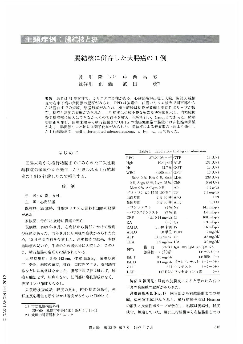
要旨 患者は61歳女性で,カリエスの既往がある.心窩部痛が出現し入院.胸部X線検査で右中下葉の葉間膜の肥厚がみられ,PPDは強陽性.注腸バリウム検査で回盲部から右結腸曲までの短縮,憩室形成がみられ,横行結腸は粘膜が萎縮し炎症性ポリープが散在,狭窄と高度の短縮がみられた.上行結腸は辺縁不整な極端な狭窄像を示し,内視鏡検査で狭窄部に挿入はできなかったので鉗子を挿入,生検を行い,Group 5であった.結腸切除術を施行.回腸末端から横行結腸までUl-Ⅱsの潰瘍瘢痕帯で腸壁には非乾酪肉芽腫があり,腸間膜リンパ節には硝子化巣がみられた.腸結核による瘢痕帯の上皮より発生した上行結腸癌で,well differentiated adenocarcinoma,s,ly2,v0,n0であった.
A 61 year-old woman who had a previous history of pelvic caries was admitted to our hospital with epigastralgia as her chief complaint.
Chest x-ray examination revealed hypertrophy of right major fissure. This was because of pleuritis. PPD reaction, also, was severe and positive. Barium meal studies showed diverticulum-like bulge, ulcer and contraction of right side colon, mucosal atrophy with many inflammatory polyps with transverse colon. There was a remarkably stenotic segment where the marginal wall was irregular in the ascending colon. Endoscopic biopsy taken from the stenosis revealed carcinoma. Right hemicolectomy with transverse colectomy was performed. Histological examination revealed a widely scarred area with ulceration and non-caseous granuloma in the right-side and transverse colons.
Histology also revealed a hyalinized tubercle in a mesenteric lymph node. It may be concluded that the carcinoma of the ascending colon originated from regenerating epithelium in an area scarred by tuberculosis. Carcinoma was classified histologically as well differentiated adenocarcinoma involving the serosa. Lymph node metastasis was negative.
Copyright © 1987, Igaku-Shoin Ltd. All rights reserved.