Japanese
English
- 有料閲覧
- Abstract 文献概要
- 1ページ目 Look Inside
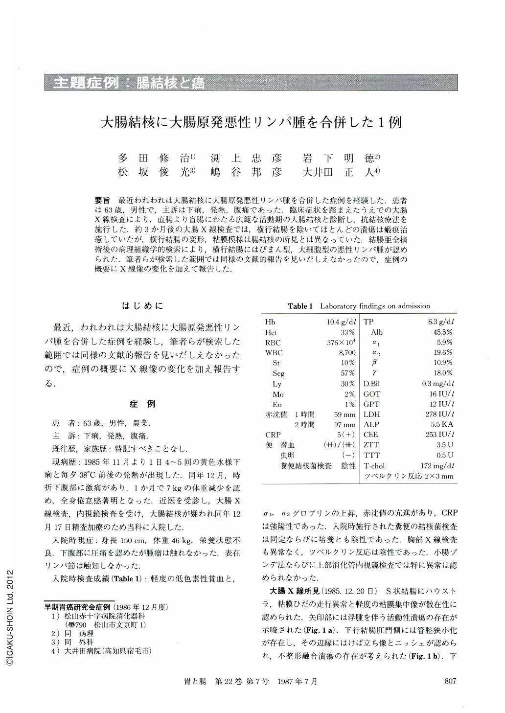
要旨 最近われわれは大腸結核に大腸原発悪性リンパ腫を合併した症例を経験した.患者は63歳,男性で,主訴は下痢,発熱,腹痛であった.臨床症状を踏まえたうえでの大腸X線検査により,直腸より盲腸にわたる広範な活動期の大腸結核と診断し,抗結核療法を施行した.約3か月後の大腸X線検査では,横行結腸を除いてほとんどの潰瘍は瘢痕治癒していたが,横行結腸の変形,粘膜模様は腸結核の所見とは異なっていた.結腸亜全摘術後の病理組織学的検索により,横行結腸にはびまん型,大細胞型の悪性リンパ腫が認められた.筆者らが検索した範囲では同様の文献的報告を見いだしえなかったので,症例の概要にX線像の変化を加えて報告した.
A 63 year-old man was admitted to the hospital on December 17, 1985, with complaints of diarrhea, fever, and abdominal pain. Laboratory findings revealed severe inflammatory signs (Table 1). Barium enema (Fig. 1a, 2a, 3a) and colonoscopy (Fig. 4a, b) showed the characteristic deformity and ulcers of colonic tuberculosis. We diagnosed the illness as colonic tuberculosis and antituberculous drugs were used. Slight improvement of his symptoms was gained but it seemed to be resistant to the therapy (Fig. 5). About three months after the initial examination, barium enema (Fig. 1b, 3b) showed the characteristics of tuberculosis except for the transverse colon. Although most active ulcers had been healed, a hose pipe stenosis and irregular ulcers remained in the transverse colon (Fig. 2b). These changes were different from the characteristic deformity of colonic tuberculosis. As his symptoms were not improved, subtotal colectomy was performed (May 6, 1986).
The operative specimen showed the shortening of the descending and ascending colon, and a large mass in the transverse colon. The histologic examination showed some almost healed ulcer scars of Ul-Ⅱ or Ul-Ⅲ and two small granulomas in the entire resected colon except for the transverse colon (Fig. 8a, b). The histoiogy of the tumor in the transverse colon showed the diffuse infiltration of large atypical lymphocytes in the entire thickness of the colonic wall (Fig. 9). The histologic findlngs were compatible with an intestinal tuberculosis in the healing stage associated with a primary malignant lymphoma in the colon.
We could not find out the same report as this case in the literature. We reported this case mainly from the radiological point of view.

Copyright © 1987, Igaku-Shoin Ltd. All rights reserved.


