Japanese
English
- 有料閲覧
PPV購入案内
1,320円 (1,200円+税10%) こちらは、781ページから785ページの文献です。購入には医書.jp本会員登録が必要です。
会員登録完了後に改めて上記「購入サイトへ」を選択してください。
または「購入サイトへ」選択後に「購入ログイン」、「新規会員登録」の順に進んでください。
会員登録について詳しくはをご確認ください。
- Abstract 文献概要
- 1ページ目 Look Inside
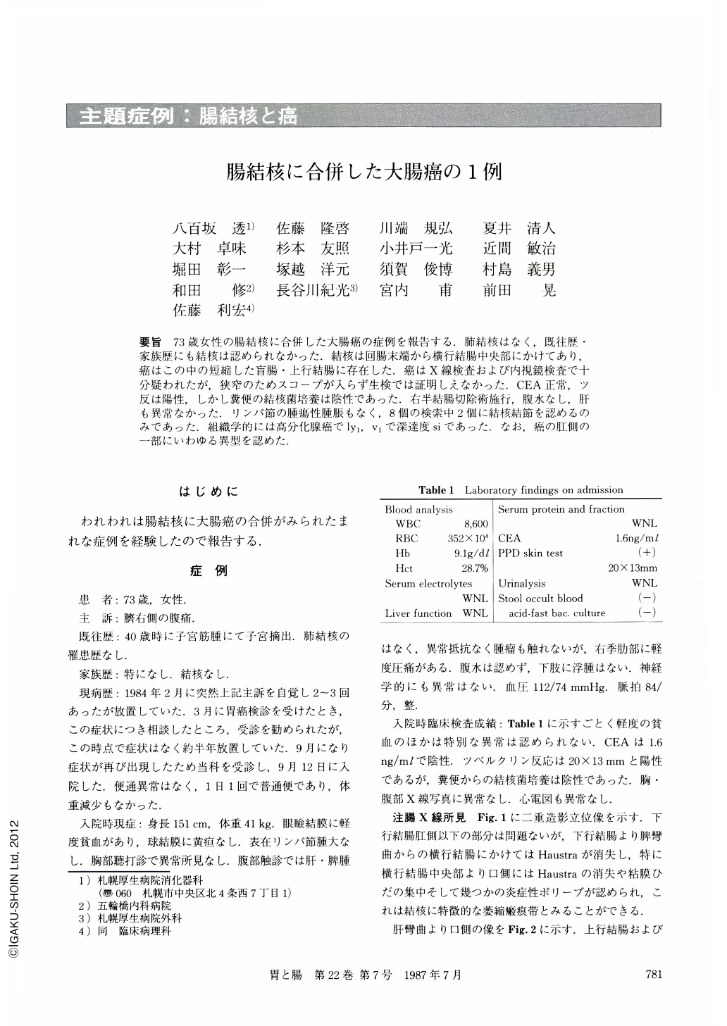
要旨 73歳女性の腸結核に合併した大腸癌の症例を報告する.肺結核はなく,既往歴・家族歴にも結核は認められなかった.結核は回腸末端から横行結腸中央部にかけてあり,癌はこの中の短縮した盲腸・上行結腸に存在した.癌はX線検査および内視鏡検査で十分疑われたが,狭窄のためスコープが入らず生検では証明しえなかった.CEA正常,ツ反は陽性,しかし糞便の結核菌培養は陰性であった.右半結腸切除術施行,腹水なし,肝も異常なかった.リンパ節の腫瘍性腫脹もなく,8個の検索中2個に結核結節を認めるのみであった.組織学的には高分化腺癌でly1,v1で深達度siであった.なお,癌の肛側の一部にいわゆる異型を認めた.
A 73 year-old woman with right para-umbilical pain was referred to our hospital for detailed examination of the colon. Neither she nor her family had any type of tuberculosis in their medical histories. Physical and laboratory examinations (Table 1) revealed no abnormalities but she had slight anemia. CEA was negative. PPD skin test was postive (20×13 mm). Acid-fast bacilli culture from her stool was negative.
Radiological examinations (Fig. 1, 2) revealed a narrowing with some irregular niches and polypoids at the shortened cecum and ascending colon. The terminal ileum and transverse colon showed atrophic mucosal pattern with some converging folds and inflammatory polyps. These radiological findings led to the diagnosis of colonic carcinoma accompanied by intestinal tuberculosis.
Endoscopic examinations (Fig. 3) could not prove carcinoma because of the impossibility of inserting the scope as far as the narrowed ascending colon. The transverse colon on the anal side of the carcinoma showed some converging folds and inflammatory polyps, which were diagnosed as tuberculosis.
The surgical specimen (Fig. 4) and its histology (Fig. 6b, 7a) showed typical intestinal tuberculosis extending from the terminal ileum to the mid-transverse colon. The carcinoma was in the shortened cecum and the ascending colon (Fig. 4, 5). It was 6.5×4.0 cm in size, and invaded the serosa (si). The histological type was that of well-differentiated adenocarcinoma (Fig. 6a). There was also a dysplastic (not cancerous) part shown by histology at the anal-side margin of this carcinoma (Fig. 7b).
Copyright © 1987, Igaku-Shoin Ltd. All rights reserved.