Japanese
English
- 有料閲覧
- Abstract 文献概要
- 1ページ目 Look Inside
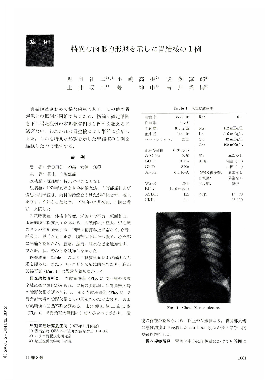
胃結核はきわめて稀な疾患であり,その他の胃疾患との鑑別が困難であるため,術前に確定診断を下し得た症例の本邦報告例は3例4)を数えるに過ぎない.われわれは胃生検により術前に診断しえた,しかも特異な形態を示した胃結核の1例を経験したので報告する.
A 29-year-old woman had complained of general malaise, loss of appetite and pain in the upper abdomen since the summer of 1974. In the beginning of December 1974. she came to us for checkup with chief complaints of nausea and occasional vomiting. X-ray examination revealed poor distensibility of the entire upper part of the corpus, mural rigidity and narrowing of the lesser curvature with a shadow defect on the greater curvature opposite to the angle. Tentative X-ray diagnosis was scirrhous carcinoma. Endoscopy disclosed slight depression of almost the entire gastric mucosa with white coating it. The depression on the greater curvature opposite to the angle was more striking. The floor of the ulcer was markedly uneven. Epithelioid tubercles in the muscularis propria were shown by biopsy. Medical management was of no avail. Gastric symptoms became intense along with melena to such a degree that we had to perform total gastrectomy.
Macroscopically the resected specimen showed an oval shallow ulcer extending from the antrum up to the cardiac region with the lesser curvature side as its center. Histologically a great number of epithelioid tubercles mixed with Langhans giant cells were seen in the muscularis propria. Some of them showed caseous necrosis. Epithelioid tubercles were also seen in the walls of venules and arterioles of the subserosa.
Although tuberculosis of the stomach has been reported since relatively old times, mostly it was a necropsied case. During the past 8 years even with increasing advance in gastric examinations, diagnosis of gastric tuberculosis, prior to either surgical operation or autopsy, has been made only in 3 cases.
The case presented here is a rare instance, diagnosed by biopsy before the operation and characterized by its short course, rapid aggravation of clinical symptoms and extensive lesions.
Copyright © 1976, Igaku-Shoin Ltd. All rights reserved.