Japanese
English
- 有料閲覧
PPV購入案内
1,320円 (1,200円+税10%) こちらは、543ページから552ページの文献です。購入には医書.jp本会員登録が必要です。
会員登録完了後に改めて上記「購入サイトへ」を選択してください。
または「購入サイトへ」選択後に「購入ログイン」、「新規会員登録」の順に進んでください。
会員登録について詳しくはをご確認ください。
- Abstract 文献概要
- 1ページ目 Look Inside
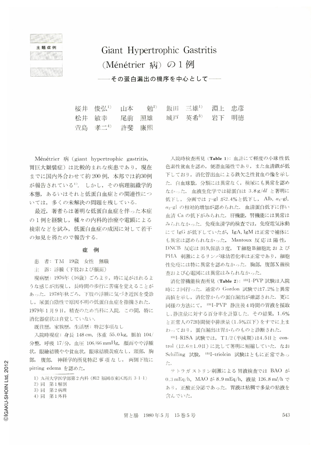
Ménétrier病(giant hypertrophic gastritis,胃巨大皺襞症)は比較的まれな疾患であり,現在までに国内外合わせて約200例,本邦では約30例が報告されている1).しかし,その病理組織学的本態,あるいはそれと低蛋白血症との関連性については,多くの未解決の問題を残している.
最近,著者らは著明な低蛋白血症を伴った本症の1例を経験し,種々の内科的治療や電顕による検索などを試み,低蛋白血症の成因に対して若干の知見を得たので報告する.
A 19 year-old female was admitted to our hospital in January 1979 because of edema of the face and lower extremities. There was no abnormal finding on physical examination except for edema of the above regions. On laboratory examination positive occult blood of feces and hypocliromic anemia were recognizecl. Proteinuria, however, was not seen. Hypoproteinemia was the only abnormal finding obtained by serum biochemical test. X-ray examination of the stomach showed huge folds in the body and innumerable elevations of coarse granular pattern in the entire stomach. On endoscopy innumerable reddish elevations were recognized on the whole gastric wall and abundant mucus was attached to the mucosa. 131I-PVP test revealed abnormally high level (7.2%), and it was proved that hypoproteinernia was due to protein loss from the gastrointestinal tract. From these results, the patient was diagnosed as having giant hypertrophic gastritis (Ménétrier's disease) associated with protein losing gastropathy.
Medical treatment, such as total parenteral nutrition, antifibrinolysis therapy (t-AMCHA) and administration of cirnetidine, was performed. Nevertheless, they were totally ineffective for protein losing and she eventually underwent total gastrectomy.
On the resected specimen, folds of the fundic gland area were remarkably thickened. Most of the folds were arranged along the longitudinal axis, some being tortuous and looking like cerebral gyri. There were innumerable sessile polyps of various size extending through the whole stomach. Histopathologically, hyperplasia of foveolar epithelium and atrophy of the pyloric and fundic glands were recognized, associated with a considerable inflammatory cell infiltrate in the mucosal stroma. On electron microscopic examination, tight junction of the epithelial cells was kept normal, but fine structures of the desquamated cells into the glandular lumen ranged from normal to highly degenerated. Plasminogen tissue activator level of the gastric mucosa was slightly high.
Serum protein level and 131I-PVP test examined a month after the operation returned to a level within a normal range.
Copyright © 1980, Igaku-Shoin Ltd. All rights reserved.