Japanese
English
- 有料閲覧
- Abstract 文献概要
- 1ページ目 Look Inside
- サイト内被引用 Cited by
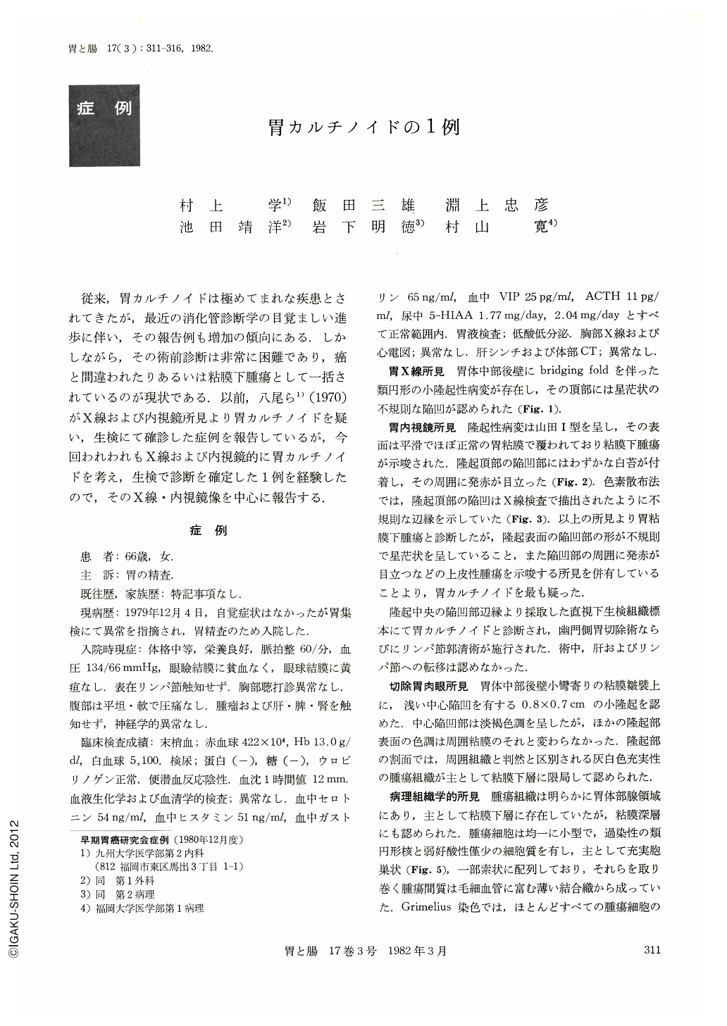
従来,胃カルチノイドは極めてまれな疾患とされてきたが,最近の消化管診断学の目覚ましい進歩に伴い,その報告例も増加の傾向にある.しかしながら,その術前診断は非常に困難であり,癌と間違われたりあるいは粘膜下腫瘍として一括されているのが現状である.以前,八尾ら1)(1970)がX線および内視鏡所見より胃カルチノイドを疑い,生検にて確診した症例を報告しているが,今回われわれもX線および内視鏡的に胃カルチノイドを考え,生検で診断を確定した1例を経験したので,そのX線・内視鏡像を中心に報告する.
A 66-year-old woman was found to have a polypoid lesion in the stomach at a mass screening of the upper G. I. tract and she was admitted to our hospital. Double contrast study of the stomach showed a small protruded mass with an irregular central depression and bridging folds in the mid-corpus. Endoscopically the broad-based polypoid tumor covered by normal mucosa was eroded and reddened on the top. Partial gastrectomy was performed under the diagnosis of gastric carcinoid by the endoscopic biopsy. The resected stomach revealed a polypoid tumor, 0.8×0.7 cm in diameters, with a central dimple in the mid-corpus. The cut surface showed a gray-white solid mass with well-defined boundaries located in the submucosa. Histologically the tumor was composed of small cells with uniform round nuclei and arranged largely in solid nests. Mitotic figures were rare. This carcinoid tumor did not give a positive argentaffin reaction, but an argyrophil. An electron microscopic appearance demonstrated numerous neurosecretory granules, measuring 150~300 nm in di-ameter, in the cytoplasms of the tumor cells. Plasma serotonin levels (54 ng/ml) and urinary 5-HIAA (1.77 mg/day, 2.04 mg/day) were within nomal limits.
Twenty-three reported cases of gastric carcinoids in Japan with a full description of radiographic and endoscopic findings were reviewed and are discussed regarding their characteristic gross appearances.

Copyright © 1982, Igaku-Shoin Ltd. All rights reserved.


