Japanese
English
- 有料閲覧
PPV購入案内
1,320円 (1,200円+税10%) こちらは、1411ページから1414ページの文献です。購入には医書.jp本会員登録が必要です。
会員登録完了後に改めて上記「購入サイトへ」を選択してください。
または「購入サイトへ」選択後に「購入ログイン」、「新規会員登録」の順に進んでください。
会員登録について詳しくはをご確認ください。
- Abstract 文献概要
- 1ページ目 Look Inside
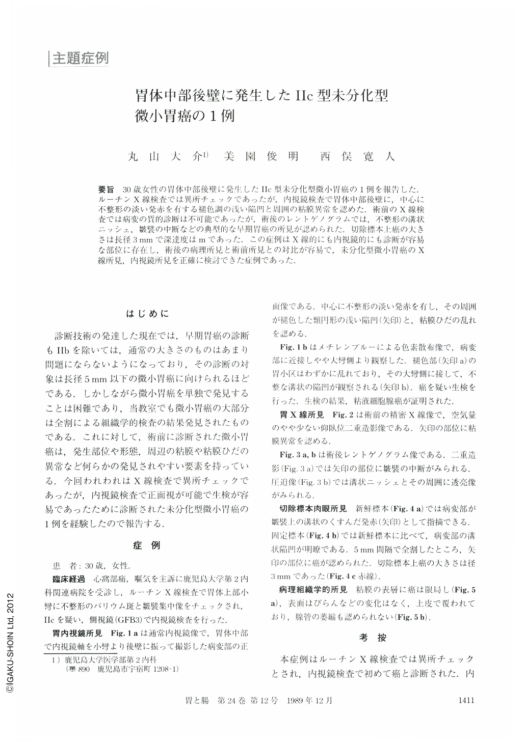
要旨 30歳女性の留体中部後壁に発生したⅡc型末分化型微小胃癌の1例を報告した.ルーチンX線検査では異所チェックであったが,内視鏡検査で胃体中部後壁に,中心に不整形の淡い発赤を有する褪色調の浅い陥凹と周囲の粘膜異常を認めた.術前のX線検査では病変の質的診断は不可能であったが,術後のレントゲノグラムでは,不整形の溝状ニッシェ,皺襞の中断などの典型的な早期胃癌の所見が認められた.切除標本上癌の大きさは長径3mmで深達度はmであった.この症例はX線的にも内視鏡的にも診断が容易な部位に存在し,術後の病理所見と術前所見との対比が容易で,未分化型微小胃癌のX線所見,内視鏡所見を正確に検討できた症例であった.
A 30-year-old woman visited our hospital complaining of epigastralgia and nausea. Endoscopic examination revealed reddish granules and a shallow depression with discoloration surrounded by abnormal mucosa (Fig. 1a, b).
Although preoperative radiography failed to demonstrate these findings, postoperative roentgenogram showed such typical findings of early gastric cancer as irregular shaped depression, granular changes, and abrupt interruption of the mucosal folds (Fig. 3 a, b).
The cancer lesion measuring 3 mm in size was confirmed in the resected stomach. Pathologically, the mucosal layer was invaded by poorly differentiated adenocarcinoma.
Copyright © 1989, Igaku-Shoin Ltd. All rights reserved.