Japanese
English
- 有料閲覧
- Abstract 文献概要
- 1ページ目 Look Inside
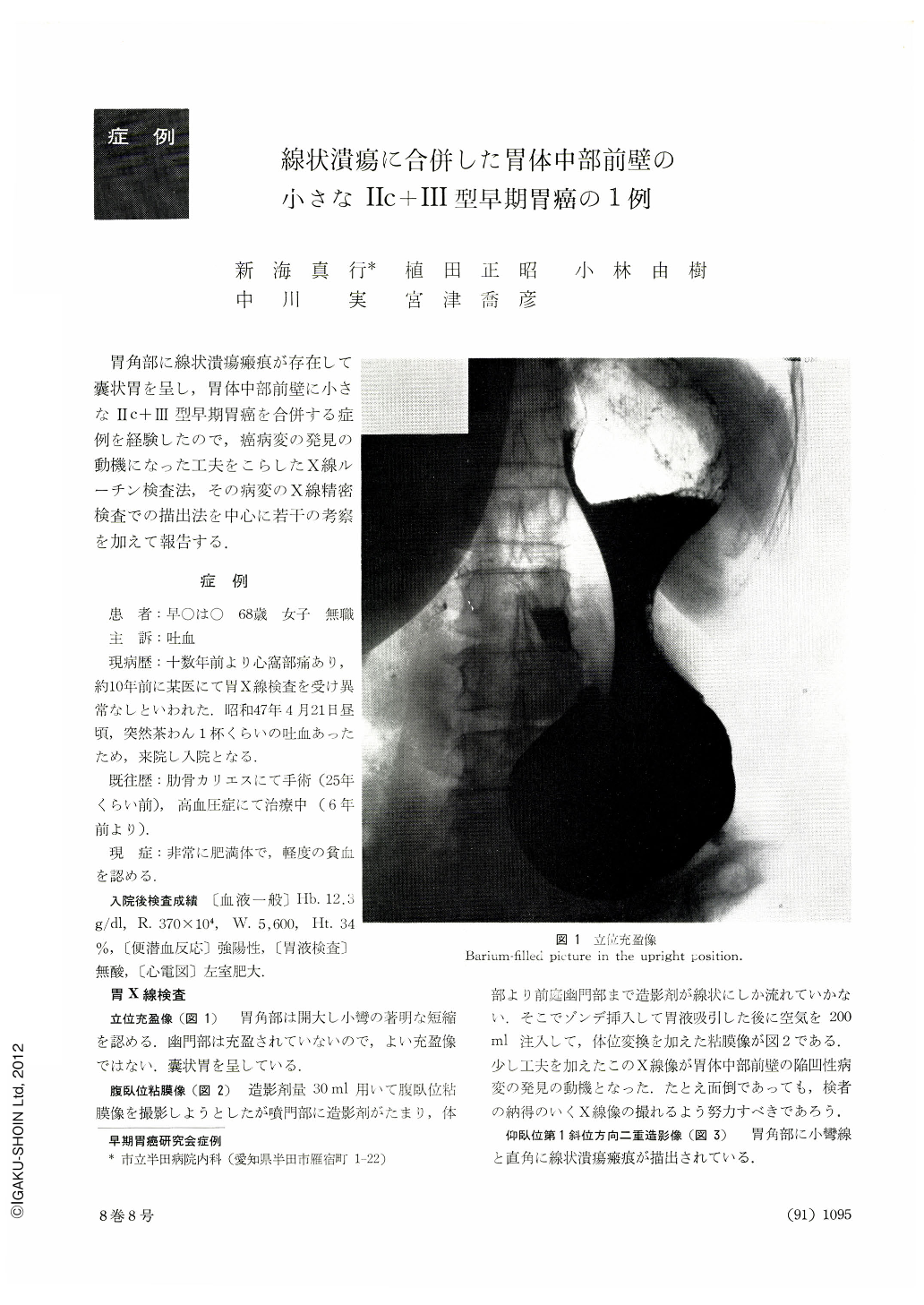
胃角部に線状潰瘍瘢痕が存在して囊状胃を呈し,胃体中部前壁に小さなⅡc+Ⅲ型早期胃癌を合併する症例を経験したので,癌病変の発見の動機になった工夫をこらしたX線ルーチン検査法,その病変のX線精密検査での描出法を中心に若干の考察を加えて報告する.
A 68-year-old woman visited our hospital with a chief complaint of hematemesis. X-ray examination revealed a deformed sack-like stomach. In routine examination we contrived to demonstrate a depressed lesion on the anterior wall of the mid-body. By detailed x-ray examination it was diagnosed as Ⅱc+Ⅲ. Pathology section of our hospital reported that biopsy was negative for cancer, but our overall clinical diagnosis was Ⅱc+Ⅲ. The patient underwent gastrectomy. The Department of Pathology of a School of Medicine we had entrusted our specimen with sent us a report with the diagnosis that the lesion on the anterior wall was a Ul-Ⅱ ulcer scar with an independent linear ulcer scar at the gastric angle. At a subsequent meeting for study, the final diagnosis was corrected: histologically the lesion on the anterior wall of the mid-body was Ⅱc+Ⅲ type early gastric cancer accompanied with a Ul-IV ulcer scar at the angle at some distance from the cancer lesion. Gastric biopsy had been positive for cancer. Clinician and pathologist must still maintain cooperative attitude. We have reperceived the fact that progress in the diagnostics of gastric diseases was made by the cooperation of roentgenologist, endoscopist, surgeon and pathologist. This case has taught us a lesson that we should always hold an attitude of humble reflection.

Copyright © 1973, Igaku-Shoin Ltd. All rights reserved.


