Japanese
English
- 有料閲覧
PPV購入案内
1,320円 (1,200円+税10%) こちらは、1303ページから1306ページの文献です。購入には医書.jp本会員登録が必要です。
会員登録完了後に改めて上記「購入サイトへ」を選択してください。
または「購入サイトへ」選択後に「購入ログイン」、「新規会員登録」の順に進んでください。
会員登録について詳しくはをご確認ください。
- Abstract 文献概要
- 1ページ目 Look Inside
- サイト内被引用 Cited by
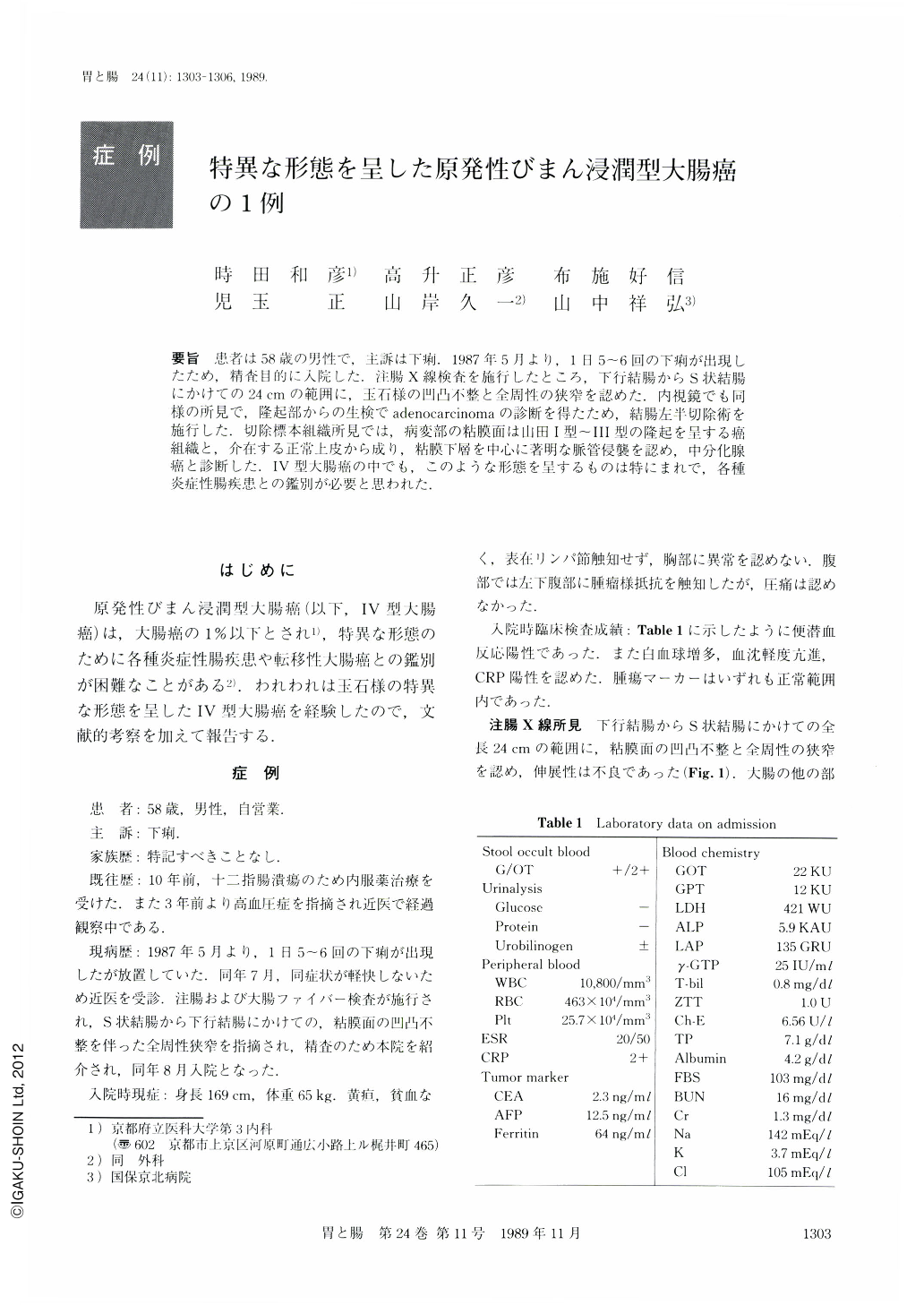
要旨 患者は58歳の男性で,主訴は下痢.1987年5月より,1日5~6回の下痢が出現したため,精査目的に入院した.注腸X線検査を施行したところ,下行結腸からS状結腸にかけての24cmの範囲に,玉石様の凹凸不整と全周性の狭窄を認めた.内視鏡でも同様の所見で,隆起部からの生検でadenocarcinomaの診断を得たため,結腸左半切除術を施行した.切除標本組織所見では,病変部の粘膜面は山田Ⅰ型~Ⅲ型の隆起を呈する癌組織と,介在する正常上皮から成り,粘膜下層を中心に著明な脈管侵襲を認め,中分化腺癌と診断した.Ⅳ型大腸癌の中でも,このような形態を呈するものは特にまれで,各種炎症性腸疾患との鑑別が必要と思われた.
A 58-year-old man was admitted to our hospital in August 1987 because of diarrhea. Stool occult blood was positive. Inflammatory bowel disease was suspected on examination of laboratory data, such as leukocytosis, increased ESR and positive CRP reaction. Barium enema showed cobblestone-like mucosa and stenosis from the descending colon to the sigmoid colon. Colonoscopic examination showed multiple polypoid lesions on the site 40 cm from the anus, but the oral side mucosa couldn't be examined because of stenosis. Biopsy specimens obtained from elevated lesions revealed adenocarcinoma, and left colectomy was performed. Macroscopic examination of the resected specimen showed irregular mucosa with multiple polypoid lesions. Elevated lesions were composed of cancer cells and massive invasion of the lymphatic vessels by cancer cells was noted in the submucosal layer by microscopic examination. It was diagnosed as moderately differentiated adenocarcinoma.
This type of colon cancer is very rare and it seems to be important to distinguish it from inflammatory bowel diseases.
Copyright © 1989, Igaku-Shoin Ltd. All rights reserved.