Japanese
English
- 有料閲覧
PPV購入案内
1,320円 (1,200円+税10%) こちらは、915ページから920ページの文献です。購入には医書.jp本会員登録が必要です。
会員登録完了後に改めて上記「購入サイトへ」を選択してください。
または「購入サイトへ」選択後に「購入ログイン」、「新規会員登録」の順に進んでください。
会員登録について詳しくはをご確認ください。
- Abstract 文献概要
- 1ページ目 Look Inside
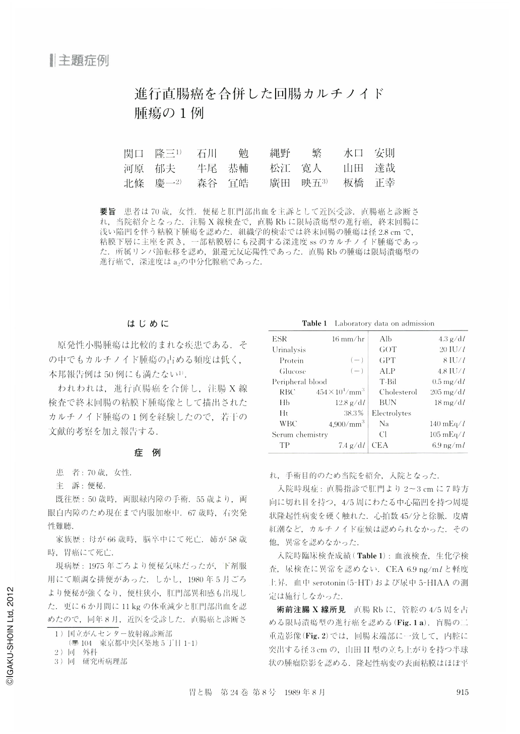
要旨 患者は70歳,女性.便秘と肛門部出血を主訴として近医受診.直腸癌と診断され,当院紹介となった.注腸X線検査で,直腸Rbに限局潰瘍型の進行癌,終末回腸に浅い陥凹を伴う粘膜下腫瘍を認めた.組織学的検索では終末回腸の腫瘍は径2.8cmで,粘膜下層に主座を置き,一部粘膜層にも浸潤する深達度ssのカルチノイド腫瘍であった.所属リンパ節転移を認め,銀還元反応陽性であった.直腸Rbの腫瘍は限局潰瘍型の進行癌で,深達度はa2の中分化腺癌であった.
A 70-year-old woman, in otherwise good health, presented with a 3-month history of constipation. Physical examination revealed type 2 advanced cancer of the rectum. Any symptoms suggestive of carcinoid syndrome were not present. Laboratory studies were negative except for slight elevation of CEA level (Table 1). The blood 5-HT and the urine 5-HIAA levels were not examined. Barium enema examination showed both type 2 advanced rectal cancer (Fig. 1) and a submucosal tumor and a shallow ulcer in the terminal ileum with otherwise smooth surface (Fig. 2). A definitive diagnosis of this tumor were not made prior to surgery. Miles' operation and right hemicolectomy with lymph node dissection were performed.
The submucosal tumor in the terminal ileum was pathologically malignant carcinoid tumor, type A+C of Soga's classification (Fig. 6 a); metastases were found in the regional lymph nodes on microscopic examination. Argyrophil (Grimelius method) and argentaffin stains (Fontana-Masson method) were strongly positive (Fig. 6 b). These findings favored the possibility of midgut origin. Type 2 advanced cancer of the rectum (Rb), moderately differentiated adenocarcinoma, was also pathologically confirmed.
Carcinoid tumor of the small intestine is quite rare in Japan. The possibility of it, however, should be entertained when a submucosal or a small polypoid tumor is seen in the small intestine, especially in the distal ileum.
Copyright © 1989, Igaku-Shoin Ltd. All rights reserved.