Japanese
English
- 有料閲覧
PPV購入案内
1,320円 (1,200円+税10%) こちらは、1237ページから1247ページの文献です。購入には医書.jp本会員登録が必要です。
会員登録完了後に改めて上記「購入サイトへ」を選択してください。
または「購入サイトへ」選択後に「購入ログイン」、「新規会員登録」の順に進んでください。
会員登録について詳しくはをご確認ください。
- Abstract 文献概要
- 1ページ目 Look Inside
- サイト内被引用 Cited by
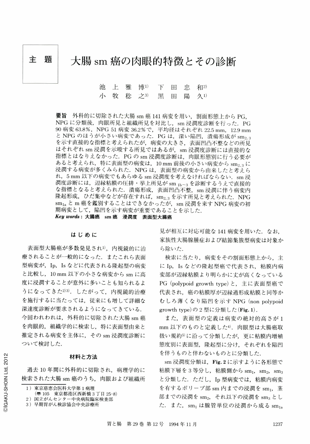
要旨 外科的に切除された大腸sm癌141病変を用い,割面形態上からPG,NPGに分類後,肉眼所見と組織所見を対比し,sm浸潤度診断を行った.PG90病変63.8%,NPG51病変36.2%で,平均径はそれぞれ22.5mm,12.9mmとNPGのほうが小さい病変であった.PGは,深い陥凹,潰瘍形成がsm2,3を示す直接的な指標と考えられたが,病変の大きさ,表面凹凸不整などの所見はそれぞれsm浸潤を示唆する所見ではあるが,sm浸潤度診断には直接的な指標とはなりえなかった.PGのsm浸潤度診断は,肉眼形態別に行う必要があると考えられ,特に表面型の病変は,10mm前後の小さい病変からsm2.3に浸潤する病変が多くみられた.NPGは,表面型の病変から由来したと考えられ,5mm以下の病変でもあらゆるsm浸潤度を考えなければならない.sm浸潤度診断には,辺縁粘膜の圧排・挙上所見がsm1b~3を診断するうえで直接的な指標となると考えられた.潰瘍形成,表面凹凸不整,sm浸潤に伴う病変内隆起形成,ひだ集中などが存在すれば,sm2,3を示す所見と考えられた.NPGsm1aとm癌を鑑別することはできなかったが,sm浸潤を来すNPG病変の初期病変として,陥凹を示す病変が重要であることを示した.
To find an association between macroscopic and microscopic findings, 141 cases of resected submucosal invasive cancer of the colorectum were studied. They were classified into two groups based on cross-section view, polypoid growth(PG) and non polypoid growth (NPG). 90 lesions(63.8%)werePG, while 51 lesions (36.2%)were NPG. NPG were smaller than PG in mean size, 12.9mm and 22.5mm respectively at the intramucosal part.
In PG, tumor size or surface irregularity suggested the existence of submucosal invasion but had no effectiveness to decide invation depth.In addition to this, deep depression or ulceration proved to be a direct indication of moderate(sm2)or severe (sm3)submucosal invasion. In PG, the depth of submucosal invasion was decided according the macroscopic appearance in each case. Superficial type PG showed moderate or severe submucosal invasion even when small in size (about 10mm)and almost all pedunculated lesions showed mild (sm1)invasion.
NPG were considered to drive from superficial type cancers. NPG, even less than 5 mm in size, showed severe submucosal invasion so often that we must consider then to have potentially every depth of submucosal invasion. In NPG, extrusion or elevation of marginal mucosa was thought to be a direct indication of mild to severe invasion. UIceration, surface irregularity, intralesional small nodes, or fold convergence usually indicated moderate to severe submucosal invasion. There's no definite difference between intramucosal and minimal invasive cancer(sm1a)in macroscopic appearance, but the lesions showing depression were thought to be important sign of the initial change that leads to submucosal invasion.
Copyright © 1994, Igaku-Shoin Ltd. All rights reserved.