Japanese
English
- 有料閲覧
PPV購入案内
1,320円 (1,200円+税10%) こちらは、1197ページから1200ページの文献です。購入には医書.jp本会員登録が必要です。
会員登録完了後に改めて上記「購入サイトへ」を選択してください。
または「購入サイトへ」選択後に「購入ログイン」、「新規会員登録」の順に進んでください。
会員登録について詳しくはをご確認ください。
- Abstract 文献概要
- 1ページ目 Look Inside
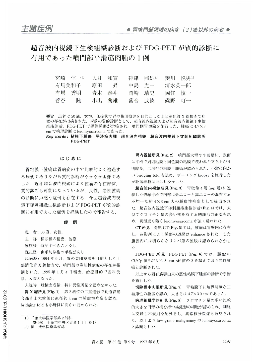
要旨 患者は50歳,女性.無症状で胃の集団検診を目的とした上部消化管X線検査で病変の存在が指摘された.術前の質的診断として,超音波内視鏡および超音波内視鏡下生検組織診断,FDG-PETで悪性腫瘍が示唆され,噴門側胃切除を施行した.腫瘍は4.7×3cmで病理診断はleiomyosarcomaであった.
A fifty-year-old woman was admitted to our department for more detailed medical evaluation and treatment of a cardiac lesion. X-ray and endoscopic examinations revealed a protruding lesion with a bridging fold on the greater curvature of cardia. EUS disclosed a tumorous lesion, 4×3 cm in diameter, of a high and low mixed echo pattern, adjacent to the mp layer. Sonopsy through EUS showed spindle shaped cells with large and chromatin rich nucleus and high atypia. The surrounding area of the lesion was enhanced by dynamic CT scan, and the Ci/Cp value of FDG-PET was 3.02 which was above the malignant cut off point of 2.0. The preoperative diagnosis of the lesion was a malignant myogenic tumor and operation was performed. The resected specimen showed the size of the lesion was 4.7×3.0 cm in diameter and histologically, leiomyosarcoma with low grade malignancy.
Copyright © 1997, Igaku-Shoin Ltd. All rights reserved.