Japanese
English
- 有料閲覧
- Abstract 文献概要
- 1ページ目 Look Inside
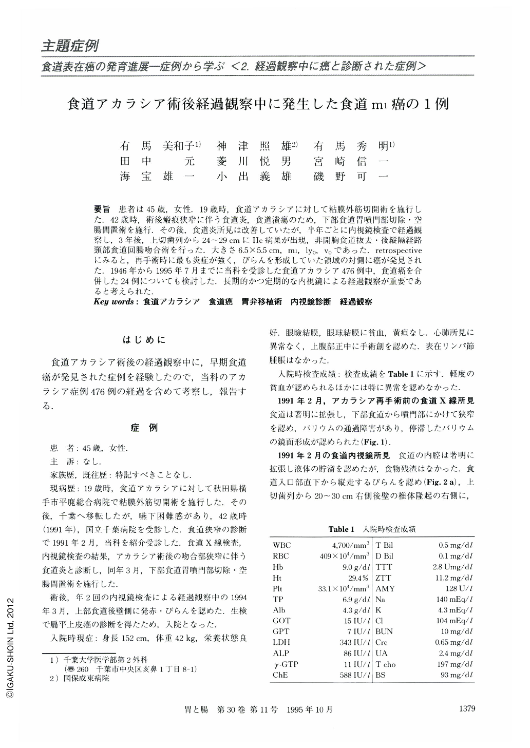
要旨 患者は45歳,女性.19歳時,食道アカラシアに対して粘膜外筋切開術を施行した.42歳時,術後瘢痕狭窄に伴う食道炎,食道潰瘍のため,下部食道胃噴門部切除・空腸間置術を施行.その後,食道炎所見は改善していたが,半年ごとに内視鏡検査で経過観察し,3年後,上切歯列から24~29cmにⅡc病巣が出現,非開胸食道抜去・後縦隔経路頸部食道回腸吻合術を行った.大きさ6.5×5.5cm,m1,ly0,v0であった.retrospectiveにみると,再手術時に最も炎症が強く,びらんを形成していた領域の対側に癌が発見された.1946年から1995年7月までに当科を受診した食道アカラシア476例中,食道癌を合併した24例についても検討した.長期的かつ定期的な内視鏡による経過観察が重要であると考えられた.
The patient was a 45-year-old female on whom lower esophageal myotomy for achalasia had been performed when she was 19 years old. At 42 years of age, lower esophagectomy and jejunum interportion had been performed for postoperative anastomotic stenosis with esophagitis. Every half year after the operation endoscopic examination was performed and esophagitis was well controlled. Three years after the second operation, a type Ⅱc esophageal cancer, 5 mm in size, appeared 24 cm from the incisors. Blunt esophagectomy and cervical esophago-ileostomy was performed. The cancer was 6.5 × 5.5 cm, m1, ly0, v0,. Endoscopically, the lesion was located at the opposite side of the most inflamed and erosive area of the esophagus observed at the second operation. On the occasion of this study 476 cases of achalasia presented at our institute from 1946 to 1995 were reviewed. Among them there were 24 cases of esophageal cancer. It is important to carry out long term and periodical endoscopic follow-up for achalasia.

Copyright © 1995, Igaku-Shoin Ltd. All rights reserved.


