Japanese
English
- 有料閲覧
- Abstract 文献概要
- 1ページ目 Look Inside
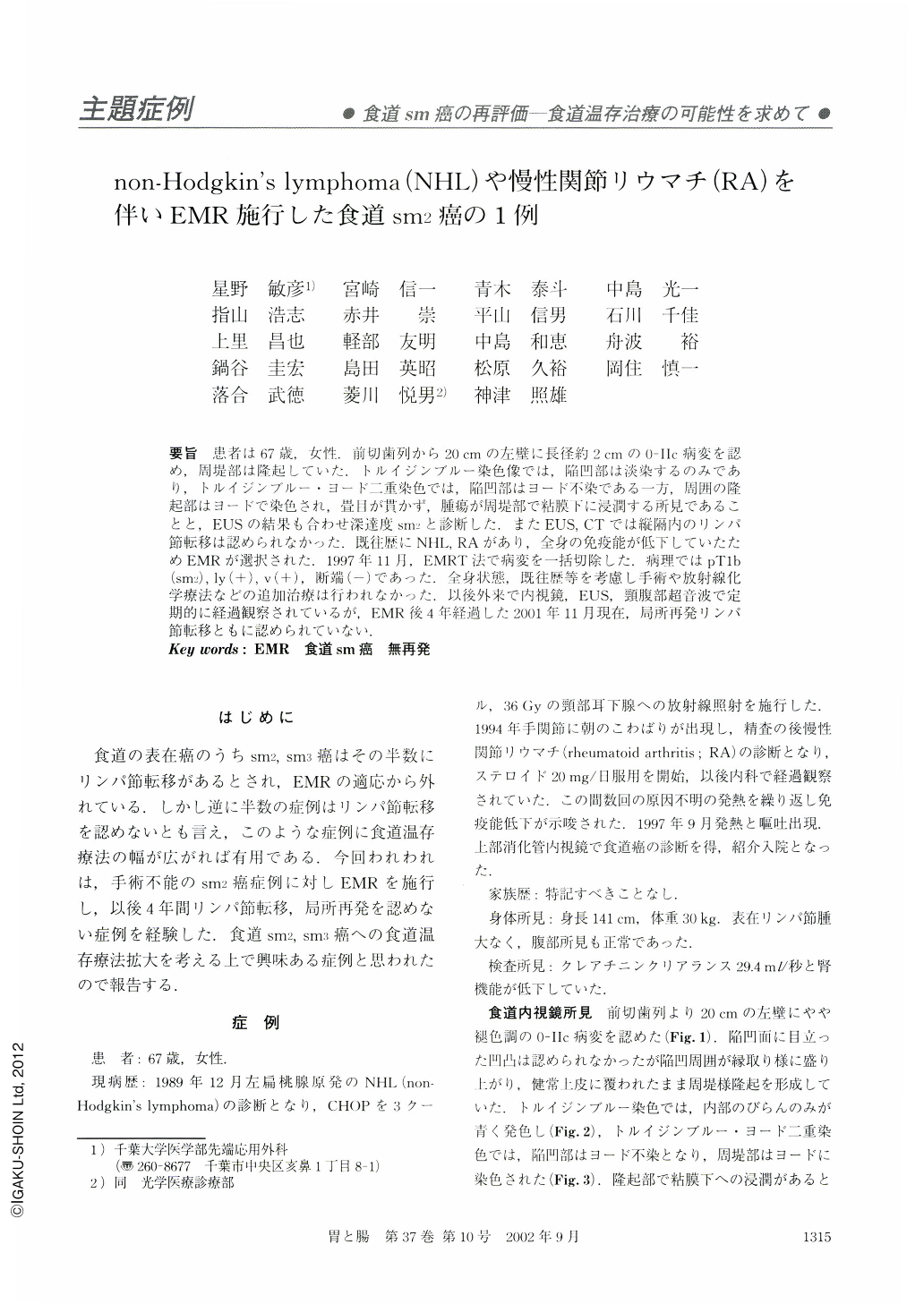
要旨 患者は67歳,女性.前切歯列から20cmの左壁に長径約2cmの0-Ⅱc病変を認め,周堤部は隆起していた.トルイジンブルー染色像では,陥凹部は淡染するのみであり,トルイジンブルー・ヨード二重染色では,陥凹部はヨード不染である一方,周囲の隆起部はヨードで染色され,畳目が貫かず,腫瘍が周堤部で粘膜下に浸潤する所見であることと,EUSの結果も合わせ深達度sm2と診断した.またEUS,CTでは縦隔内のリンパ節転移は認められなかった.既往歴にNHL,RAがあり,全身の免疫能が低下していたためEMRが選択された.1997年11月,EMRT法で病変を一括切除した.病理ではpT1b(sm2),ly(+),v(+),断端(-)であった.全身状態,既往歴等を考慮し手術や放射線化学療法などの追加治療は行われなかった.以後外来で内視鏡,EUS,頸腹部超音波で定期的に経過観察されているが,EMR後4年経過した2001年11月現在,局所再発リンパ節転移ともに認められていない.
The patient was a 67-year-old female. A flat 0-Ⅱc lesion was recognized in the esophageal wall 20 cm away from the incision. In the endoscopic examination, double staining method using iodine dye and toluidine blue was performed and it revealed that the lesion boundary was indistinct and was not stained with iodine dye, whereas a part of the lesion stained blue with toluidine dye. The invasion depth of the lesion was diagnosed as sm2, which accounted for the same diagnosis being obtained by EUS performed during the endoscopy. Neither EUS nor CT verified lymph node metastasis. Since there were NHL, Sjögren Syndrome, and RA in the patient's medical history, her general immunocompetence was weakened, so we selected a management method using EMR. The lesion was removed through EMR by the two-channel method in November, 1997. The pathological findings revealed that the lesion was pT1b (sm2), ly(+), v(+), stump(-). Considering her general conditions and medical history, neither operation nor radio-chemotherapy was performed. She has been undergoing periodical follow-ups with endoscopy, EUS, and abdominal ultrasonography on an ambulatory basis, and no local recurrence or lymph node metastasis have yet been found as of November, 2001.

Copyright © 2002, Igaku-Shoin Ltd. All rights reserved.


