Japanese
English
- 有料閲覧
PPV購入案内
1,320円 (1,200円+税10%) こちらは、267ページから274ページの文献です。購入には医書.jp本会員登録が必要です。
会員登録完了後に改めて上記「購入サイトへ」を選択してください。
または「購入サイトへ」選択後に「購入ログイン」、「新規会員登録」の順に進んでください。
会員登録について詳しくはをご確認ください。
- Abstract 文献概要
- 1ページ目 Look Inside
- サイト内被引用 Cited by
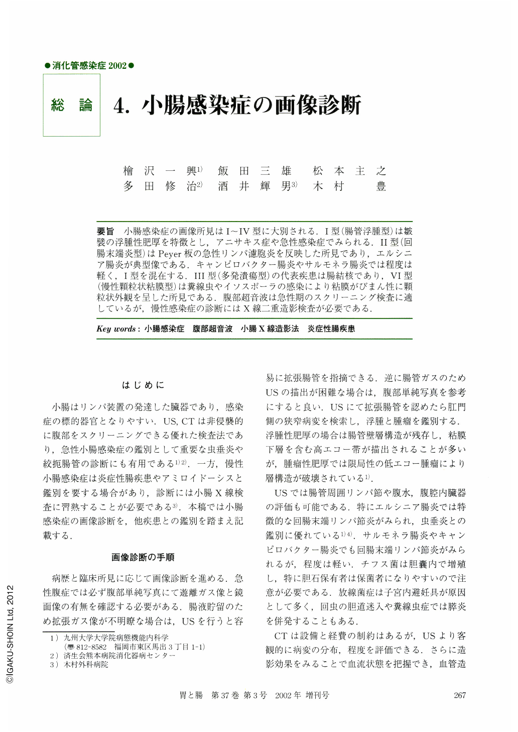
要旨 小腸感染症の画像所見はⅠ~Ⅳ型に大別される.Ⅰ型(腸管浮腫型)は皺襞の浮腫性肥厚を特徴とし,アニサキス症や急性感染症でみられる.Ⅱ型(回腸末端炎型)はPeyer板の急性リンパ濾胞炎を反映した所見であり,エルシニア腸炎が典型像である.キャンピロバクター腸炎やサルモネラ腸炎では程度は軽く,Ⅰ型を混在する.Ⅲ型(多発潰瘍型)の代表疾患は腸結核であり,Ⅵ型(慢性顆粒状粘膜型)は糞線虫やイソスポーラの感染により粘膜がびまん性に顆粒状外観を呈した所見である.腹部超音波は急性期のスクリーニング検査に適しているが,慢性感染症の診断にはX線二重造影検査が必要である.
Diagnostic findings of infectious enteritis are mainly categorized as type Ⅰ (edematous thickening of the valvulae conniventes), type Ⅱ (lymph folliculitis of the terminal ileum), type Ⅲ (multiple intestinal ulcers), and type Ⅳ (diffusely granular mucosa caused by chronic infection). Anisakiasis and Vibrio enteritis are classified into type Ⅰ. Acute infection of Campylobacter jejuni and Salmonella organisms manifest type Ⅰ and Ⅱ. Type Ⅲ is typified by intestinal tuberculosis. Strongyloidiasis and isosporiasis gradually progress to type Ⅳ. Ultrasonography is available for evaluating the severity of acute infection, whereas double-contrast radiography of the small intestine is essential for the diagnosis of chronic enteritis.
Copyright © 2002, Igaku-Shoin Ltd. All rights reserved.