Japanese
English
- 有料閲覧
- Abstract 文献概要
- 1ページ目 Look Inside
- サイト内被引用 Cited by
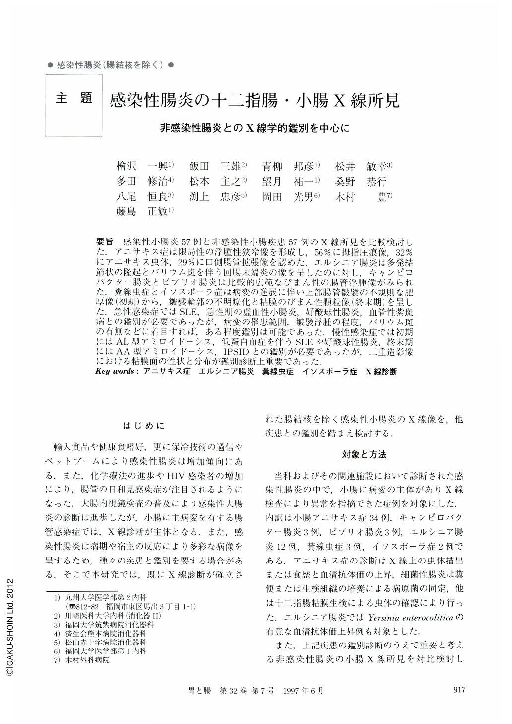
要旨 感染性小腸炎57例と非感染性小腸疾患57例のX線所見を比較検討した.アニサキス症は限局性の浮腫性狭窄像を形成し,56%に栂指圧痕像,32%にアニサキス虫体,29%に口側腸管拡張像を認めた.エルシニア腸炎は多発結節状の隆起とバリウム斑を伴う回腸末端炎の像を呈したのに対し,キャンピロバクター腸炎とビブリオ腸炎は比較的広範なびまん性の腸管浮腫像がみられた.糞線虫症とイソスポーラ症は病変の進展に伴い上部腸管皺襞の不規則な肥厚像(初期)から,皺襞輪郭の不明瞭化と粘膜のびまん性顆粒像(終末期)を呈した.急性感染症ではSLE,急性期の虚血性小腸炎,好酸球性腸炎,血管性紫斑病との鑑別が必要であったが,病変の罹患範囲,皺襞浮腫の程度,バリウム斑の有無などに着目すれば,ある程度鑑別は可能であった.慢性感染症では初期にはAL型アミロイドーシス,低蛋白血症を伴うSLEや好酸球性腸炎,終末期にはAA型アミロイドーシス,IPSIDとの鑑別が必要であったが,二重造影像における粘膜面の性状と分布が鑑別診断上重要であった.
Radiographic features of the duodenum and small intestine were retrospectively analyzed in 57 cases of infectious enteritis. These findings were also compared to those in 57 cases of non-infectious diseases, including systemic lupus erythematosus, ischemic enteritis in the acute phase, eosinophilic enteritis, Shoenlein-Henoch purpura, intestinal amyloidosis, and immunoproliferative small intestinal disease. Anisakiasis (34 cases) depicted irregularly edematous folds of the intestine, accompanied by thumbprinting in 56% of the cases and transient narrowing of the lumen with proximal bowel dilatation in 29%. Yersiniosis (12 cases) was characterized by localized terminal ileitis, presenting multiple nodules with tiny barium flecks. Infection of Vibrio parahaemolyticus (three cases) and Campylobacter jejuni (three cases) demonstrated diffusely edematous folds of the jejunum and ileum. Strongyloidiasis (three cases) and isosporiasis (two cases) initially showed irregular thickened folds of the duodenum and jejunum, and later progressed to diffusely granular mucosa and indistinct contour of the valvulae conniventes. In order to differentiate infectious enteritis from other noninfectious enteritis, it is necessary to assess the location, degree, and pattern of intestinal edema or granular mucosa on radiography of the small intestine.

Copyright © 1997, Igaku-Shoin Ltd. All rights reserved.


