Japanese
English
- 有料閲覧
- Abstract 文献概要
- 1ページ目 Look Inside
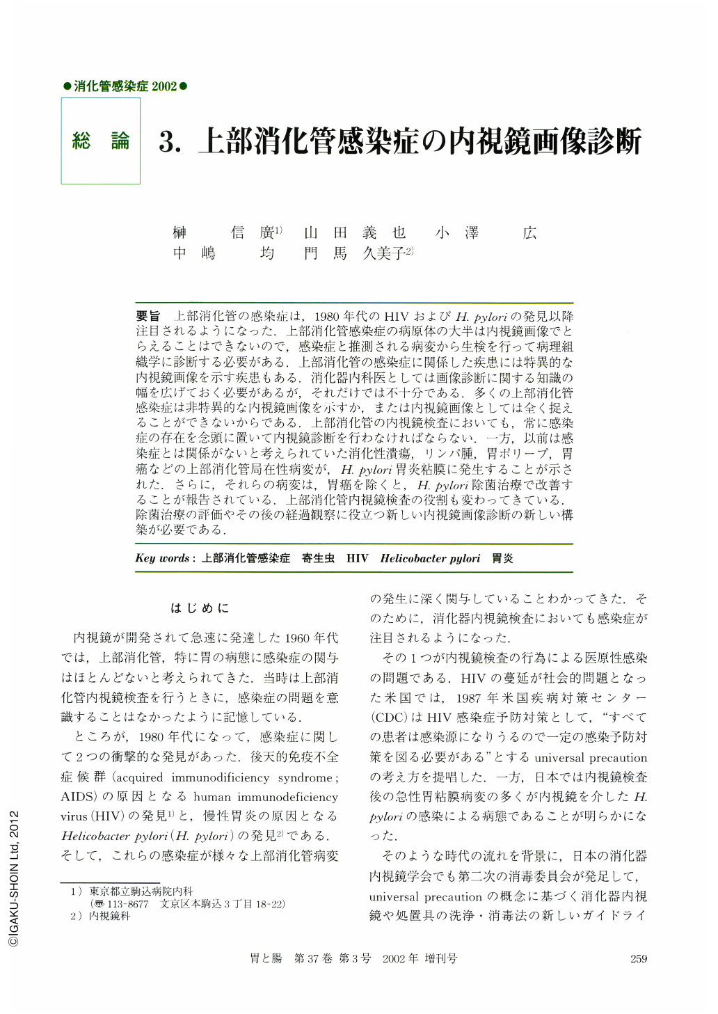
要旨 上部消化管の感染症は,1980年代のHIVおよび H. pyloriの発見以降注目されるようになった.上部消化管感染症の病原体の大半は内視鏡画像でとらえることはできないので,感染症と推測される病変から生検を行って病理組織学に診断する必要がある.上部消化管の感染症に関係した疾患には特異的な内視鏡画像を示す疾患もある.消化器内科医としては画像診断に関する知識の幅を広げておく必要があるが,それだけでは不十分である.多くの上部消化管感染症は非特異的な内視鏡画像を示すか,または内視鏡画像としては全く捉えることができないからである.上部消化管の内視鏡検査においても,常に感染の存在を念頭に置いて内視鏡診断を行わなければならない.一方,以前は感染症とは関係がないと考えられていた消化性潰瘍,リンパ腫,胃ポリープ,胃癌などの上部消化管局在性病変が,H. pylori胃炎粘膜に発生することが示された.さらに,それらの病変は,胃癌を除くと,H. pylori除菌治療で改善することが報告されている.上部消化管内視鏡検査の役割も変わってきている.除菌治療の評価やその後の経過観察に役立つ新しい内視鏡画像診断の新しい構築が必要である.
Infectious diseases of the upper-gastrointestinal (UGI) tract have been the object of deep interest since the 1980s when the human immunodeficiency virus (HIV) and Helicobacter pylori (H. pylori) were discovered. Almost all pathogens infecting the UGI tract were too small to observe by endoscopy alone. Histological diagnosis using biopsy specimens taken from the infected area is necessary to ensure a correct diagnosis. Only a few lesions, such as the esophageal Cytomegalovirus ulcer in AIDS patients, are easily diagnosed from their characteristic endoscopic findings. Therefore, when an endoscopic examination is performed for patients with an infectious disorder, biopsies should be taken even when the disorder is non-specific and even from endoscopically normal areas.
On the other hand, H. pylori is changing the role of endoscopic examination for the UGI tract. Many localized lesions located in the UGI tract; such as, peptic ulcer, MALT lymphoma, hyperplastic polyp and carcinoma are present in H. pylori-gastritis mucosa. Furthermore, these lesions except for carcinoma often improve after successful H. pylori eradication therapy. New criteria are needed for assessing the effect of eradication therapy and for endoscopic follow-up after treatment.

Copyright © 2002, Igaku-Shoin Ltd. All rights reserved.


