Japanese
English
- 有料閲覧
PPV購入案内
1,320円 (1,200円+税10%) こちらは、327ページから332ページの文献です。購入には医書.jp本会員登録が必要です。
会員登録完了後に改めて上記「購入サイトへ」を選択してください。
または「購入サイトへ」選択後に「購入ログイン」、「新規会員登録」の順に進んでください。
会員登録について詳しくはをご確認ください。
- Abstract 文献概要
- 1ページ目 Look Inside
- 参考文献 Reference
- サイト内被引用 Cited by
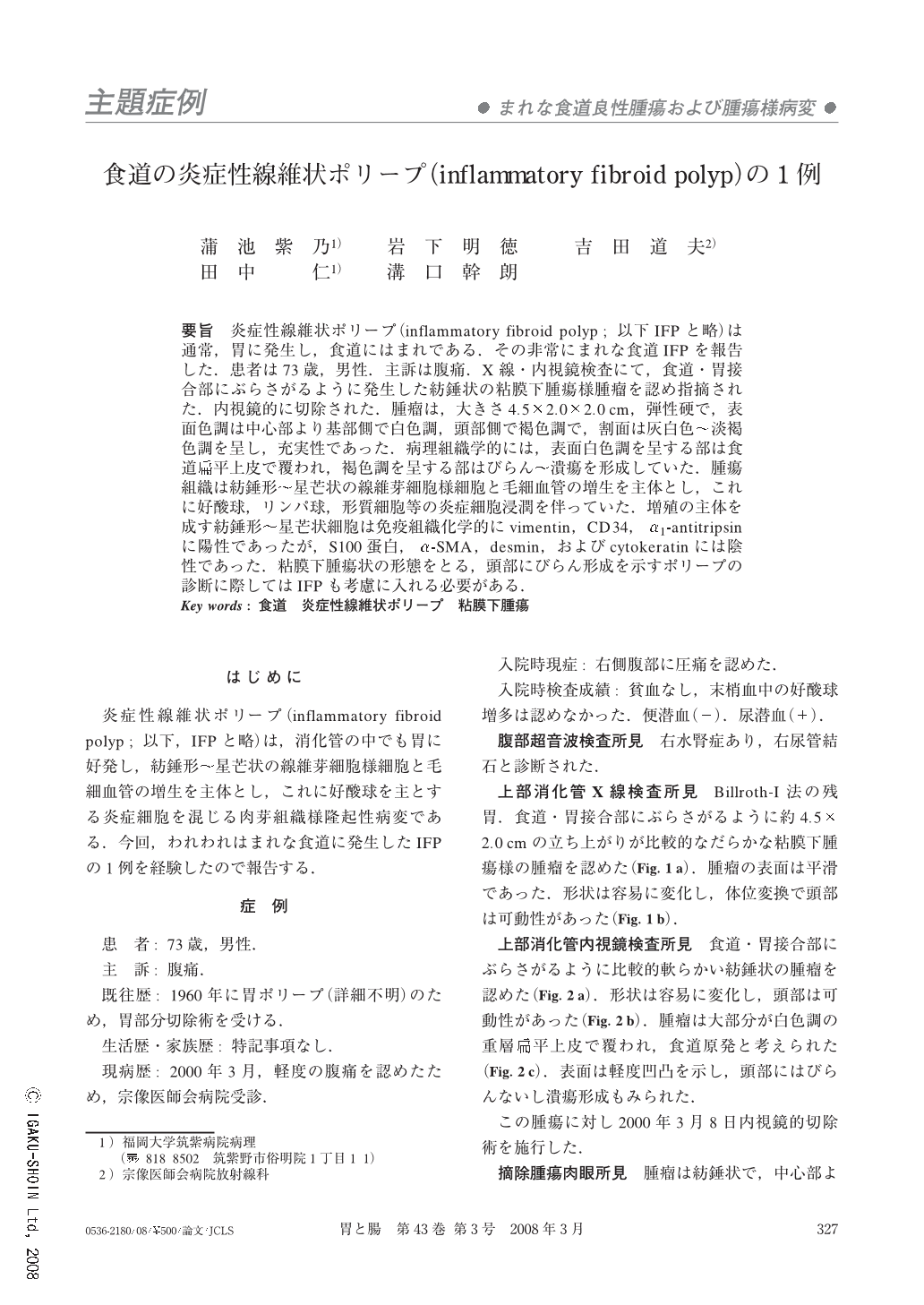
要旨 炎症性線維状ポリープ(inflammatory fibroid polyp;以下IFPと略)は通常,胃に発生し,食道にはまれである.その非常にまれな食道IFPを報告した.患者は73歳,男性.主訴は腹痛.X線・内視鏡検査にて,食道・胃接合部にぶらさがるように発生した紡錘状の粘膜下腫瘍様腫瘤を認め指摘された.内視鏡的に切除された.腫瘤は,大きさ4.5×2.0×2.0cm,弾性硬で,表面色調は中心部より基部側で白色調,頭部側で褐色調で,割面は灰白色~淡褐色調を呈し,充実性であった.病理組織学的には,表面白色調を呈する部は食道扁平上皮で覆われ,褐色調を呈する部はびらん~潰瘍を形成していた.腫瘍組織は紡錘形~星芒状の線維芽細胞様細胞と毛細血管の増生を主体とし,これに好酸球,リンパ球,形質細胞等の炎症細胞浸潤を伴っていた.増殖の主体を成す紡錘形~星芒状細胞は免疫組織化学的にvimentin,CD34,α1-antitripsinに陽性であったが,S100蛋白,α-SMA,desmin,およびcytokeratinには陰性であった.粘膜下腫瘍状の形態をとる,頭部にびらん形成を示すポリープの診断に際してはIFPも考慮に入れる必要がある.
We experienced a rare esophageal inflammatory fibroid polyp (IFP), it is generally seen in the stomach.
A 73-year old man was admitted to our hospital because of abdominal pain. Gastroroentgenography and endoscopy demonstrated a hanged spindle-shaped lesion, resembling a submucosal tumor in the esophago-gastric junction. Endoscopic resection was performed. The tumor was 4.5 cm×2.0 cm×2.0 cm in size, elastic hard. The colour of the surface was white from the middle to the base of tumor, otherwise it showed brown in the head. Divided view of the resected specimen revealed that the pale brown tumor was solid. Histopathologically, white surface are was covered with esophageal squamous mucosa, and brown colour area was formed as erosions or ulcers. The tumor included predominantly spindle-or asteroid-shaped fibroblast-like cells and showed increased capillary vessels, with inflammatory cell infiltration by eosinophils, lymphocytes, and plasma cells. Immunohistochemical tests showed the tumor to be positive for vimentin, CD34, and α1-antitrypsin, but negative for S100 protein,α-SMA, desmin, and cytokeratin on spindle-or asteroid-shaped cells, which were the major proliferated component of the tumor. IFP of the esophagus should be included as a differential diagnosis when it showed resembling a submucosal tumor with an erosion in the head.
Copyright © 2008, Igaku-Shoin Ltd. All rights reserved.