Japanese
English
- 有料閲覧
PPV購入案内
1,320円 (1,200円+税10%) こちらは、705ページから710ページの文献です。購入には医書.jp本会員登録が必要です。
会員登録完了後に改めて上記「購入サイトへ」を選択してください。
または「購入サイトへ」選択後に「購入ログイン」、「新規会員登録」の順に進んでください。
会員登録について詳しくはをご確認ください。
- Abstract 文献概要
- 1ページ目 Look Inside
- 参考文献 Reference
- サイト内被引用 Cited by
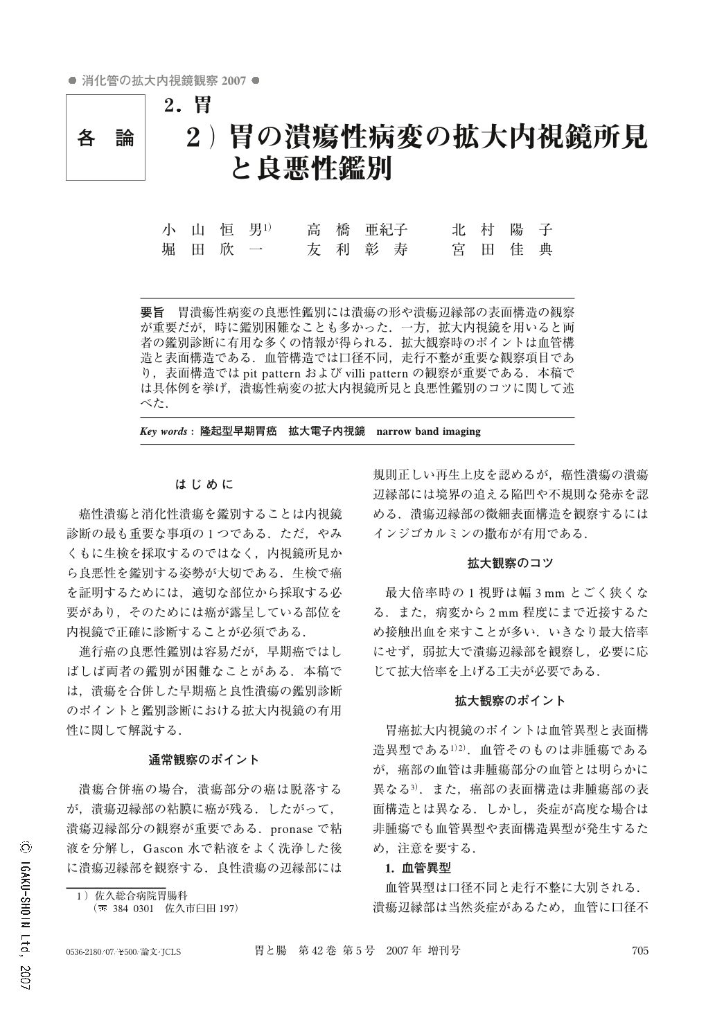
要旨 胃潰瘍性病変の良悪性鑑別には潰瘍の形や潰瘍辺縁部の表面構造の観察が重要だが,時に鑑別困難なことも多かった.一方,拡大内視鏡を用いると両者の鑑別診断に有用な多くの情報が得られる.拡大観察時のポイントは血管構造と表面構造である.血管構造では口径不同,走行不整が重要な観察項目であり,表面構造ではpit patternおよびvilli patternの観察が重要である.本稿では具体例を挙げ,潰瘍性病変の拡大内視鏡所見と良悪性鑑別のコツに関して述べた.
The differential diagnosis of benign and malignant ulcers has been regarded as important. However, it was sometimes difficult to differentiated these ulcers using conventional endoscopy.
Magnified endoscopy with the NBI system showed not only the surface pattern but also the vascular pattern of the mucosa. The check points of NBI magnified endoscopy for the diagnosis of ulcerative lesions were described in this paper.
The surface pattern was divided into pit and villi. And the irregularity and density should be checked. The points to observe in the vessels were caliber change and irregularity of shape.
Two benign ulcers and three malignant ulcers were shown in this paper, and the findings of NBI magnified endoscopy were very useful for the differential diagnosis between benign and malignant ulcers.
Copyright © 2007, Igaku-Shoin Ltd. All rights reserved.