Japanese
English
- 有料閲覧
PPV購入案内
1,320円 (1,200円+税10%) こちらは、589ページから595ページの文献です。購入には医書.jp本会員登録が必要です。
会員登録完了後に改めて上記「購入サイトへ」を選択してください。
または「購入サイトへ」選択後に「購入ログイン」、「新規会員登録」の順に進んでください。
会員登録について詳しくはをご確認ください。
- Abstract 文献概要
- 1ページ目 Look Inside
- 参考文献 Reference
- サイト内被引用 Cited by
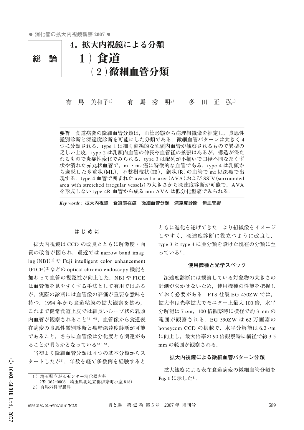
要旨 食道病変の微細血管分類は,血管形態から病理組織像を推定し,良悪性鑑別診断と深達度診断を可能にした分類である.微細血管パターンは大きく4つに分類される.type1は細く直線的な乳頭内血管が観察されるもので異型の乏しい上皮,type2は乳頭内血管の伸長や血管径の拡張はあるが,構造が保たれるもので炎症性変化でみられる.type3は配列が不揃いで口径不同な糸くず状や潰れた赤丸状血管で,m1・m2癌に特徴的な血管である.type4は乳頭から逸脱した多重状(ML),不整樹枝状(IB),網状(R)の血管でm2以深癌で出現する.type4血管で囲まれたavascular area(AVA)およびSSIV(surrounded area with stretched irregular vessels)の大きさから深達度診断が可能で,AVAを形成しないtype4R血管から成るnon-AVAは低分化型癌でみられる.
Assessment of microvascular patterns on magnifying endoscopy was useful for evaluating benign and malignant superficial esophageal lesions and for estimating the depth of cancer invasion, as well as for predicting histopathological features. Microvascular patterns on magnifying endoscopy were classified into 4 types. Type1 was characterized by thin, linear capillaries in the subepithelial papilla and was generally seen in normal mucosa. Type2 was characterized by distended, dilated vessels with variations such as branched or spiral enlargement. The structure of capillaries in the subepithelial papilla was preserved, and their arrangement was relatively regular. Type2 was generally seen in inflammatory lesions. Type3 was characterized by destruction of vessels in the subepithelial papilla, spiral vessels with an irregular caliber, and crushed vessels with red spots. The arrangement of the vessels was irregular. Type3 was generally seen in m1 or m2 cancers. Type4 was characterized by multi-layered, irregularly branched, reticular vessels with an irregular caliber. Type4 was generally seen in cancers with m2 or deeper invasion. The sizes of avascular areas (AVAs) surrounded by type4 vessels could be used to predict the depth of tumor invasion. Type4 R lesions without the formation of AVA (non-AVA) often showed invasion patterns typical of poorly differentiated carcinomas.
Copyright © 2007, Igaku-Shoin Ltd. All rights reserved.