Japanese
English
- 有料閲覧
PPV購入案内
1,320円 (1,200円+税10%) こちらは、809ページから812ページの文献です。購入には医書.jp本会員登録が必要です。
会員登録完了後に改めて上記「購入サイトへ」を選択してください。
または「購入サイトへ」選択後に「購入ログイン」、「新規会員登録」の順に進んでください。
会員登録について詳しくはをご確認ください。
- Abstract 文献概要
- 1ページ目 Look Inside
- 参考文献 Reference
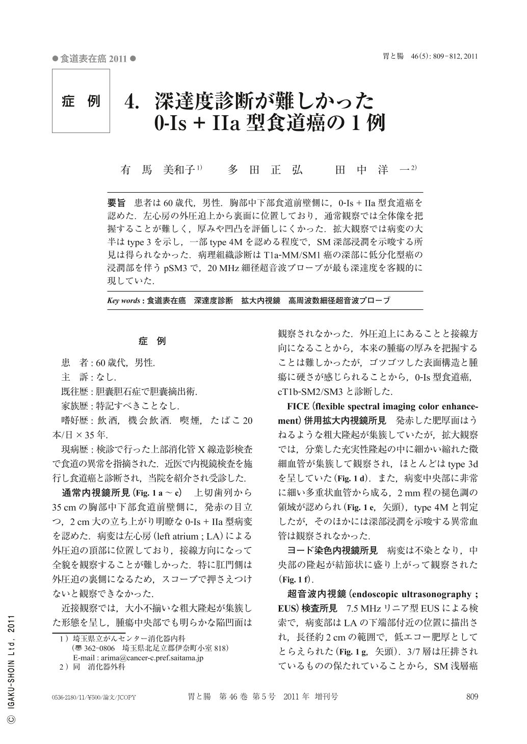
要旨 患者は60歳代,男性.胸部中下部食道前壁側に,0-Is+IIa型食道癌を認めた.左心房の外圧迫上から裏面に位置しており,通常観察では全体像を把握することが難しく,厚みや凹凸を評価しにくかった.拡大観察では病変の大半はtype 3を示し,一部type 4Mを認める程度で,SM深部浸潤を示唆する所見は得られなかった.病理組織診断はT1a-MM/SM1癌の深部に低分化型癌の浸潤部を伴うpSM3で,20MHz細径超音波プローブが最も深達度を客観的に現していた.
A type 0-Is+IIa esophageal cancer was noted in the anterior wall of the lower thoracic esophagus of a 60-year-old man. The tumor was compressed by, and located behind, the left atrium. It was difficult to view the entire tumor on conventional endoscopy. The thickness and surface characteristics of the tumor were difficult to assess. On magnifying endoscopy, most of the lesion showed type 3 microvascular pattern, and type 4M vessels were seen in only part of the lesion. There were no findings suggesting deep submucosal invasion. Histopathological examination showed a pSM3, associated with poorly differentiated carcinoma invading the region from the T1a-MM/SM1. 20MHz miniature ultrasound probe showed the depth of invasion most objectively.
Copyright © 2011, Igaku-Shoin Ltd. All rights reserved.