Japanese
English
- 有料閲覧
- Abstract 文献概要
- 1ページ目 Look Inside
- 参考文献 Reference
- サイト内被引用 Cited by
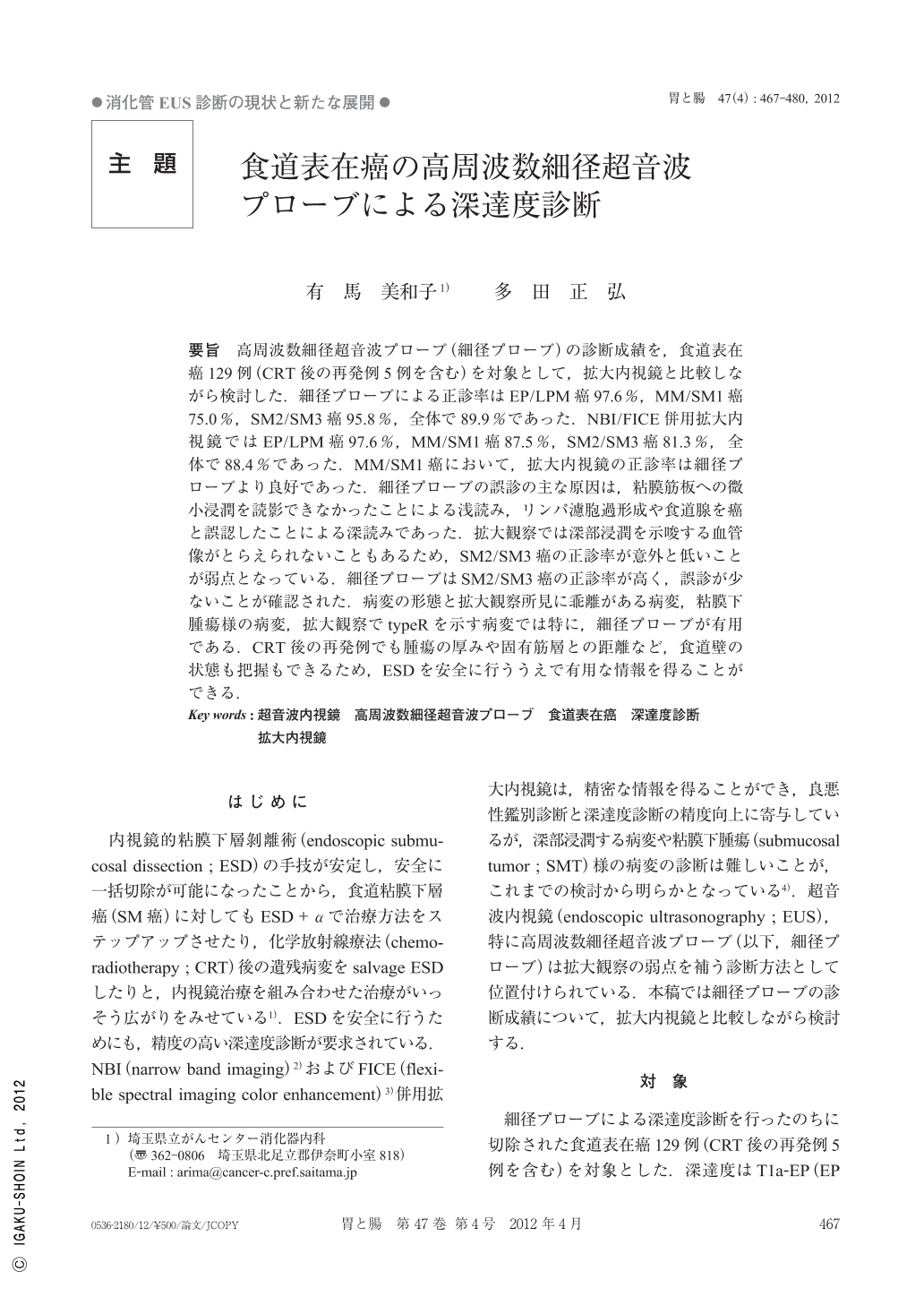
要旨 高周波数細径超音波プローブ(細径プローブ)の診断成績を,食道表在癌129例(CRT後の再発例5例を含む)を対象として,拡大内視鏡と比較しながら検討した.細径プローブによる正診率はEP/LPM癌97.6%,MM/SM1癌75.0%,SM2/SM3癌95.8%,全体で89.9%であった.NBI/FICE併用拡大内視鏡ではEP/LPM癌97.6%,MM/SM1癌87.5%,SM2/SM3癌81.3%,全体で88.4%であった.MM/SM1癌において,拡大内視鏡の正診率は細径プローブより良好であった.細径プローブの誤診の主な原因は,粘膜筋板への微小浸潤を読影できなかったことによる浅読み,リンパ濾胞過形成や食道腺を癌と誤認したことによる深読みであった.拡大観察では深部浸潤を示唆する血管像がとらえられないこともあるため,SM2/SM3癌の正診率が意外と低いことが弱点となっている.細径プローブはSM2/SM3癌の正診率が高く,誤診が少ないことが確認された.病変の形態と拡大観察所見に乖離がある病変,粘膜下腫瘍様の病変,拡大観察でtypeRを示す病変では特に,細径プローブが有用である.CRT後の再発例でも腫瘍の厚みや固有筋層との距離など,食道壁の状態も把握もできるため,ESDを安全に行ううえで有用な情報を得ることができる.
We compared diagnostic results obtained with a high-frequency miniature ultrasonic probe(mini probe)with those obtained by magnifying endoscopy in 129 cases with superficial esophageal cancer, including 5 with recurrence after CRT(chemoradiotherapy). The correct rate of diagnosis with a mini probe was 97.6% for EP/LPM cancers, 75.0% for MM/SM1cancers, 95.8% for SM2/SM3 cancers, and 89.9% overall. The correct rate of diagnosis on magnifying endoscopy combined with NBI and FICE was 97.6% for EP/LPM cancers, 87.5% for MM/SM1 cancers, 81.3% for SM2/SM3 cancers, and 88.4% overall. For MM/SM1 cancers, the correct rate of diagnosis was higher with magnifying endoscopy than with a mini probe. The main reasons for misdiagnosis of the depth of invasion by a mini probe were underestimation caused by inability to visualize microinvasion to the MM and overestimation caused by the misdiagnosis of lympholicle hyperplasia and esophageal glands as cancer. Because vascular patterns suggesting deep invasion sometimes could not be detected on magnifying endoscopy, the rate of correctly diagnosing SM2/SM3 cancers was lower than expected, suggesting that this is a drawback. A mini probe has a high rate of correctly diagnosing SM2/SM3 cancers, confirming a low rate of misdiagnosis. A mini probe is particularly useful for the diagnosis of lesions with morphologic characteristics that deviate considerably from magnifying endoscopic findings, submucosal tumor-like lesions, and type R(reticular)lesions on magnifying endoscopy. Even in patients with recurrence after CRT, a mini probe enabled tumor thickness, the distance from the MP layer, and the status of the esophageal wall to be evaluated, thereby providing useful information for the safe performance of endoscopic submucosal dissection.

Copyright © 2012, Igaku-Shoin Ltd. All rights reserved.


