Japanese
English
- 有料閲覧
PPV購入案内
1,320円 (1,200円+税10%) こちらは、1675ページから1687ページの文献です。購入には医書.jp本会員登録が必要です。
会員登録完了後に改めて上記「購入サイトへ」を選択してください。
または「購入サイトへ」選択後に「購入ログイン」、「新規会員登録」の順に進んでください。
会員登録について詳しくはをご確認ください。
- Abstract 文献概要
- 1ページ目 Look Inside
- 参考文献 Reference
- サイト内被引用 Cited by
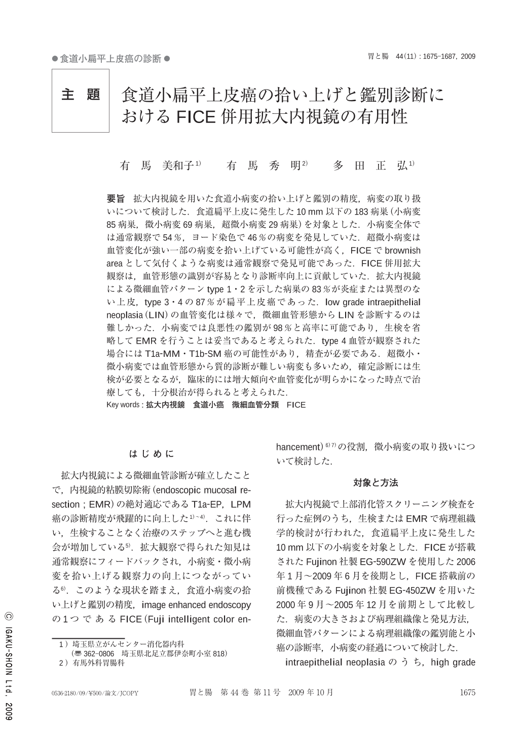
要旨 拡大内視鏡を用いた食道小病変の拾い上げと鑑別の精度,病変の取り扱いについて検討した.食道扁平上皮に発生した10mm以下の183病巣(小病変85病巣,微小病変69病巣,超微小病変29病巣)を対象とした.小病変全体では通常観察で54%,ヨード染色で46%の病変を発見していた.超微小病変は血管変化が強い一部の病変を拾い上げている可能性が高く,FICEでbrownish areaとして気付くような病変は通常観察で発見可能であった.FICE併用拡大観察は,血管形態の識別が容易となり診断率向上に貢献していた.拡大内視鏡による微細血管パターンtype1・2を示した病巣の83%が炎症または異型のない上皮,type3・4の87%が扁平上皮癌であった.low grade intraepithelial neoplasia(LIN)の血管変化は様々で,微細血管形態からLINを診断するのは難しかった.小病変では良悪性の鑑別が98%と高率に可能であり,生検を省略してEMRを行うことは妥当であると考えられた.type4血管が観察された場合にはT1a-MM・T1b-SM癌の可能性があり,精査が必要である.超微小・微小病変では血管形態から質的診断が難しい病変も多いため,確定診断には生検が必要となるが,臨床的には増大傾向や血管変化が明らかになった時点で治療しても,十分根治が得られると考えられた.
We examined whether magnifying endoscopy is useful for the screening, differential diagnosis, and treatment of small squamous cell carcinomas of the esophagus. A total of 183 lesions(85 small lesions, 69 micro lesions, and 29 supermicro lesions)10 mm or less in diameter that arose in the squamous cell epithelium of the esophagus were studied. Of all small lesions, 54% were detected on conventional endoscopy, and 46% were detected under iodine staining. Many supermicro lesions detected on endoscopy were associated with marked vascular changes. Lesions recognized as a brownish area on image enhanced endoscopy with Fujinon intelligent color enhancement(FICE)could be detected on conventional endoscopy. Magnifying endoscopy with FICE facilitated the assessment of vascular patterns, thereby contributing to improved diagnostic accuracy. On magnifying endoscopy, 83% of lesions with type 1 and 2 microvascular patterns were judged to be inflammation or with no evidence of atypia, and 87% of lesions with type 3 and 4 microvascular patterns were squamous cell carcinomas. Low grade intraepithelial neoplasia was associated with various vascular changes and was difficult to diagnose on the basis of microvascular patterns. A very high proportion of small lesions(98%)could be differentially diagnosed as benign or malignant lesions, suggesting that EMR could be performed without biopsy. Lesions with type 4 vessels may invade the muscularis mucosae or submucosa and should therefore be examined in detail. Many supermicro lesions and micro lesions are difficult to diagnose qualitatively on the basis of vascular patterns. Biopsy is therefore required for a definite diagnosis. Clinically, however, we believe that curative treatment is possible if lesions are resected when they show slight enlargement or distinct changes in vascular patterns.
Copyright © 2009, Igaku-Shoin Ltd. All rights reserved.