Japanese
English
- 有料閲覧
PPV購入案内
2,200円 (2,000円+税10%) こちらは、1075ページから1082ページの文献です。購入には医書.jp本会員登録が必要です。
会員登録完了後に改めて上記「購入サイトへ」を選択してください。
または「購入サイトへ」選択後に「購入ログイン」、「新規会員登録」の順に進んでください。
会員登録について詳しくはをご確認ください。
- Abstract 文献概要
- 1ページ目 Look Inside
- 参考文献 Reference
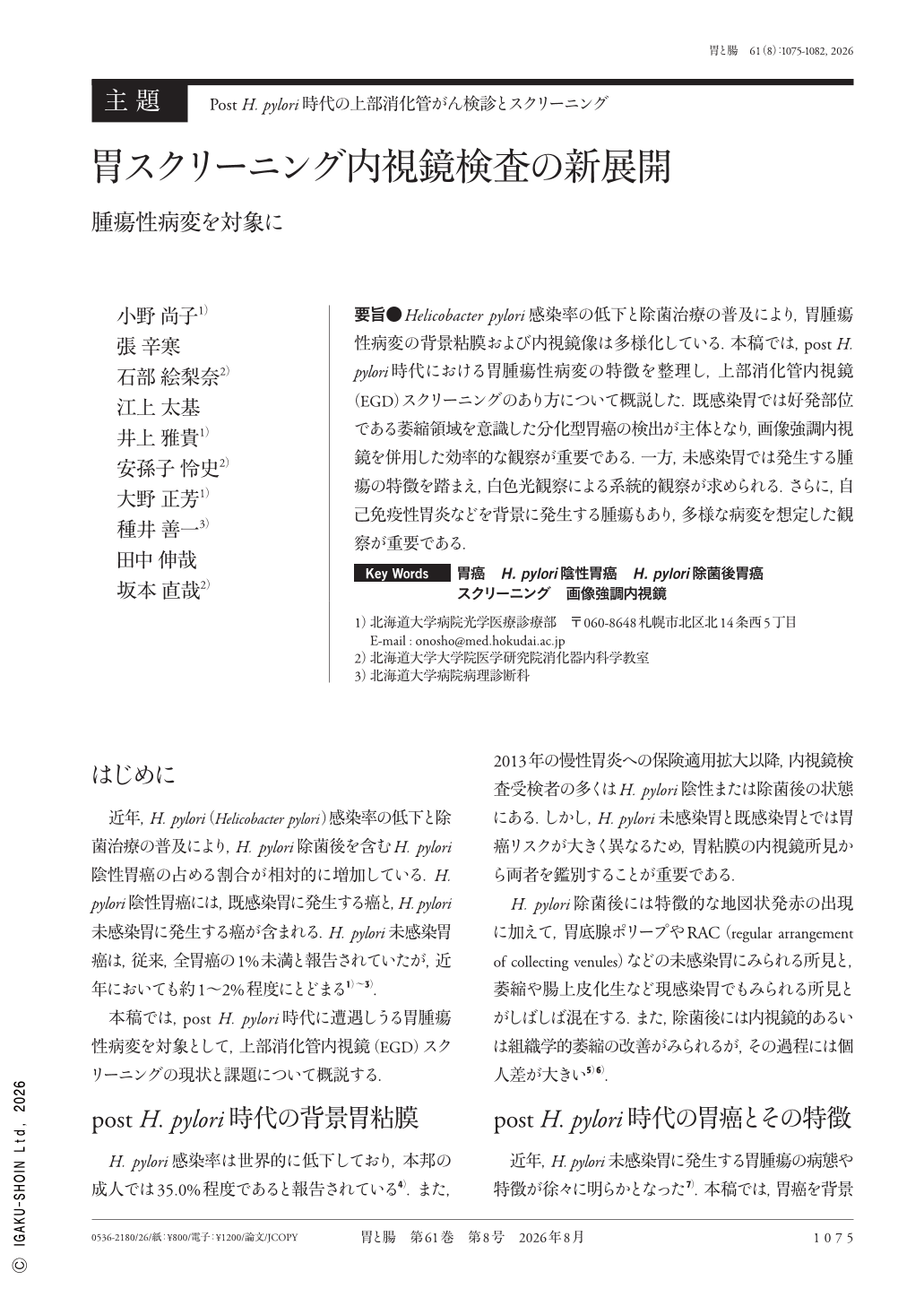
要旨●Helicobacter pylori感染率の低下と除菌治療の普及により,胃腫瘍性病変の背景粘膜および内視鏡像は多様化している.本稿では,post H. pylori時代における胃腫瘍性病変の特徴を整理し,上部消化管内視鏡(EGD)スクリーニングのあり方について概説した.既感染胃では好発部位である萎縮領域を意識した分化型胃癌の検出が主体となり,画像強調内視鏡を併用した効率的な観察が重要である.一方,未感染胃では発生する腫瘍の特徴を踏まえ,白色光観察による系統的観察が求められる.さらに,自己免疫性胃炎などを背景に発生する腫瘍もあり,多様な病変を想定した観察が重要である.
With declining prevalence of Helicobacter pylori infection and the widespread implementation of eradication therapy, the endoscopic appearance of gastric neoplastic lesions, as well as the surrounding gastric mucosa, have become increasingly diverse. This review summarizes the features of such lesions associated with prior H. pylori infection and discusses current approaches for upper gastrointestinal screening. In stomachs with prior H. pylori infection, screening primarily focuses on detecting differentiated-type gastric cancer arising in atrophic areas, particularly within high-risk regions. In addition, efficient observation using image-enhanced endoscopy is also essential. Conversely, the systematic examination of stomachs without H. pylori infection includes white-light imaging on the basis of the characteristic features of tumors that develop in this setting. Furthermore, neoplastic lesions may also arise in specific pathological backgrounds, including autoimmune gastritis ; therefore, careful observation with broad awareness of the diverse lesion types is crucial.
Copyright © 2026, Igaku-Shoin Ltd. All rights reserved.