Japanese
English
症例
4D-CTを撮像した毛細血管性腎血管腫の1例
A case of capillary renal hemangioma imaged by 4D-CT
白石 明日香
1
,
松井 裕輔
2
,
宗友 一晃
1
,
児島 克英
1
,
檜垣 文代
1
,
都地 友紘
3
,
平木 隆夫
2
Asuka Shiraishi
1
1岡山大学病院 放射線科
2岡山大学学術研究院医歯薬学域 放射線医学
3岡山赤十字病院 病理診断科
1Department of Radiology Okayama University Hospital
キーワード:
腎血管腫
,
毛細血管性腎血管腫
,
4D-CT
Keyword:
腎血管腫
,
毛細血管性腎血管腫
,
4D-CT
pp.197-200
発行日 2023年2月10日
Published Date 2023/2/10
DOI https://doi.org/10.18888/rp.0000002266
- 有料閲覧
- Abstract 文献概要
- 1ページ目 Look Inside
- 参考文献 Reference
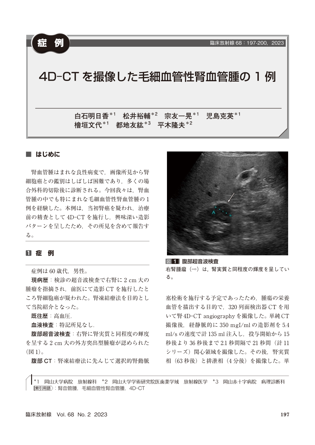
腎血管腫はまれな良性病変で,画像所見から腎細胞癌との鑑別はしばしば困難であり,多くの場合外科的切除後に診断される。今回我々は,腎血管腫の中でも特にまれな毛細血管性腎血管腫の1例を経験した。本例は,当初腎癌を疑われ,治療前の精査として4D-CTを施行し,興味深い造影パターンを呈したため,その所見を含めて報告する。
Renal hemangiomas are rare benign lesions that are difficult to be diagnosed radiologically and often diagnosed after surgical resection. We report a case of capillary renal hemangioma, a rare type of renal hemangioma, which was imaged by 4D-CT and diagnosed by CT-guided biopsy. 4D-CT showed early contrast enhancement at the periphery of the lesion that progressed over time. The peripheral-dominant enhancement persisted on the renal parenchymal phase and washed out on the excretory phase.

Copyright © 2023, KANEHARA SHUPPAN Co.LTD. All rights reserved.


