Japanese
English
- 有料閲覧
- Abstract 文献概要
- 1ページ目 Look Inside
- 参考文献 Reference
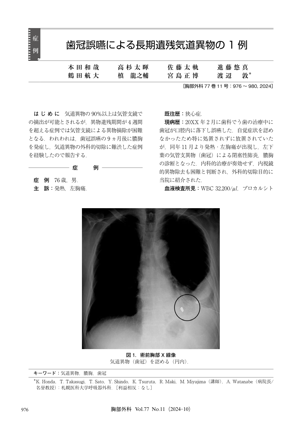
はじめに 気道異物の90%以上は気管支鏡での摘出が可能とされるが,異物遺残期間が4週間を超える症例では気管支鏡による異物摘除が困難となる.われわれは,歯冠誤嚥の9ヵ月後に膿胸を発症し,気道異物の外科的切除に難渋した症例を経験したので報告する.
A 76-year-old man presented after aspiration of a crown during dental treatment. He had no immediate symptoms;therefore, the crown was not thoroughly examined at the time of the event. The patient developed high fever and chest pain and sought medical attention, 9 months later. Chest computed tomography (CT) revealed the dental crown located in the left B10 bronchus. The patient was diagnosed with pyothorax and obstructive pneumonia and was referred to our hospital for surgical resection. We attempted partial lung resection;however, pleurisy prevented adequate palpation. Therefore, we used X-ray fluoroscopy to complete the surgery. This case report highlights the challenges associated with detection of long-standing foreign bodies in the airway. Careful preparation, including X-ray fluoroscopy and preoperative bronchoscopy, is essential for successful management.

© Nankodo Co., Ltd., 2024


