Japanese
English
- 有料閲覧
- Abstract 文献概要
- 1ページ目 Look Inside
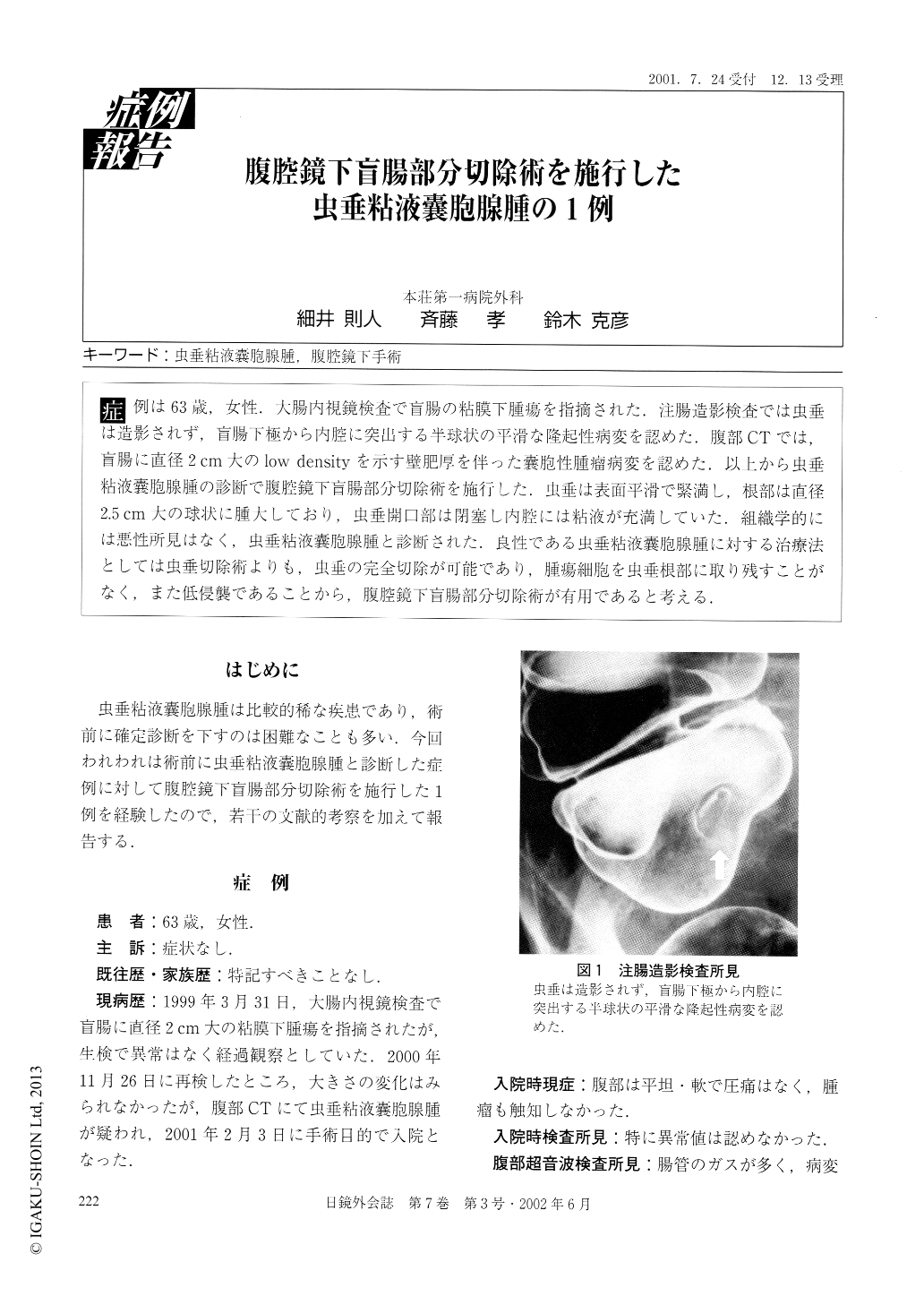
症例は63歳,女性.大腸内視鏡検査で盲腸の粘膜下腫瘍を指摘された.注腸造影検査では虫垂は造影されず,盲腸下極から内腔に突出する半球状の平滑な隆起性病変を認めた.腹部CTでは,盲腸に直径2cm大のlow densityを示す壁肥厚を伴った嚢胞性腫瘤病変を認めた.以上から虫垂粘液嚢胞腺腫の診断で腹腔鏡下盲腸部分切除術を施行した.虫垂は表面平滑で緊満し,根部は直径2.5cm大の球状に腫大しており,虫垂開口部は閉塞し内腔には粘液が充満していた.組織学的には悪性所見はなく,虫垂粘液嚢胞腺腫と診断された.良性である虫垂粘液嚢胞腺腫に対する治療法としては虫垂切除術よりも,虫垂の完全切除が可能であり,腫瘍細胞を虫垂根部に取り残すことがなく,また低侵襲であることから,腹腔鏡下盲腸部分切除術が有用であると考える.
A 63 year-old woman was pointed out as having a submucosal tumor of the cecum by a colonofiberscopy. Barium enema study did not visualize the appendix. but revealed a hemispheric smooth lesion protruding into the lumen from the lower pole of the cecum. Abdominal CT scan revealed a cystic lesion in the cecum 2 cm in diameter with wall thickening. Mucinous cystadenoma of the appendix was diagnosed and laparoscopic partial excision of the cecum was performed. The appendix was tense and had a smooth surface, with a spherically swollen root with a diameter of 2.5 cm.

Copyright © 2002, JAPAN SOCIETY FOR ENDOSCOPIC SURGERY All rights reserved.


