Japanese
English
- 有料閲覧
- Abstract 文献概要
- 1ページ目 Look Inside
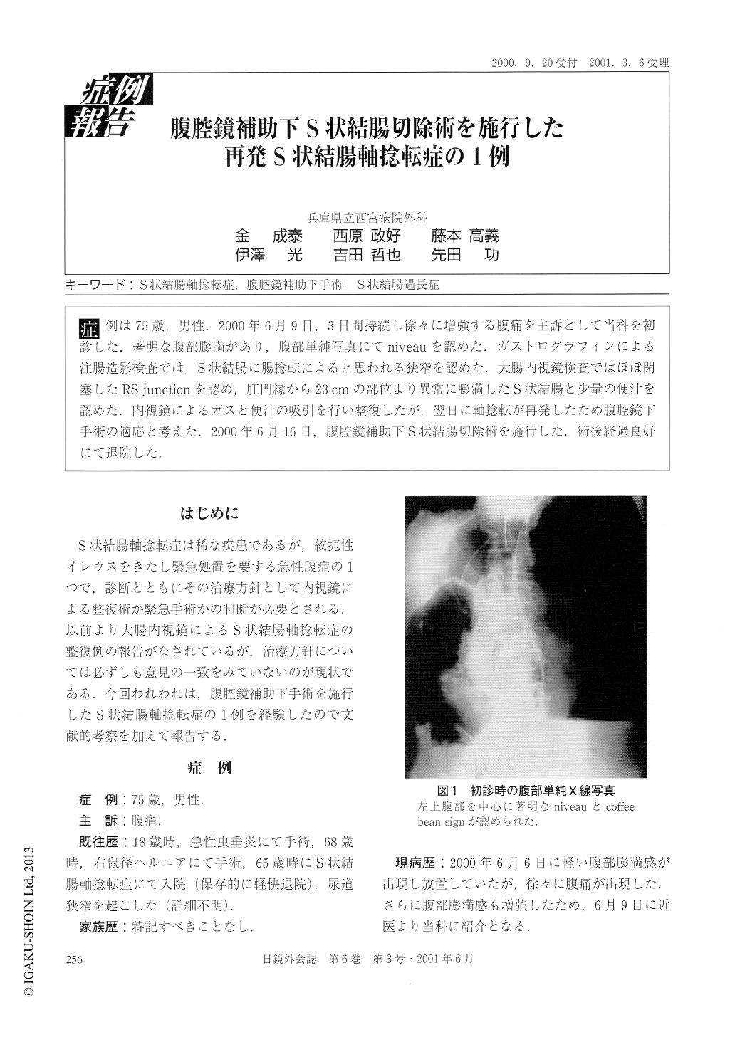
症例は75歳,男性,2000年6月9日,3日間持続し徐々に増強する腹痛を主訴として当科を初診した.著明な腹部膨満があり,腹部単純写真にてniveauを認めた.ガストログラフィンによる注腸造影検査では,S状結腸に腸捻転によると思われる狭窄を認めた.大腸内視鏡検査ではほぼ閉塞したRS junctionを認め,肛門縁から23cmの部位より異常に膨満したS状結腸と少量の便汁を認めた.内視鏡によるガスと便汁の吸引を行い整復したが,翌日に軸捻転が再発したため腹腔鏡下手術の適応と考えた.2000年6月16日,腹腔鏡補助下S状結腸切除術を施行した.術後経過良好にて退院した.
A 75-year-old male was seen at the clinic because of the abdominal pain that continued for three days. The bowel was distended mainly on the left side and there was niveau abdominal X-ray. Although there was no tumor, gastrographin-enema examination revealed a stenosis due to volvulus of the sigmoid colon. Colonoscopic examination showed an obstructive recto-sigmoid junction and unusually distended sigmoid colon, 23 cm from the anal verge. There were feces at the oral side from the stenotic lesion. The abdominal distention was reduced by suction of the air and the feces using colonoscopy.

Copyright © 2001, JAPAN SOCIETY FOR ENDOSCOPIC SURGERY All rights reserved.


