Japanese
English
- 有料閲覧
- Abstract 文献概要
- 1ページ目 Look Inside
- 参考文献 Reference
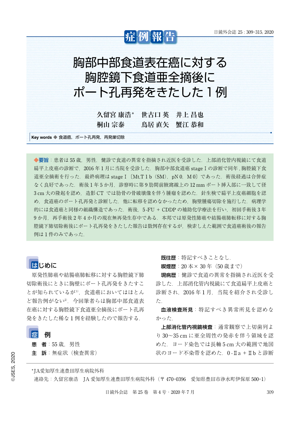
◆要旨:患者は55歳,男性.健診で食道の異常を指摘され近医を受診した.上部消化管内視鏡にて食道扁平上皮癌の診断で,2016年1月に当院を受診した.胸部中部食道癌stageⅠの診断で同年,胸腔鏡下食道亜全摘術を行った.最終病理はstageⅠ〔Mt,T1b(SM),pN0,M0〕であった.術後経過は合併症なく良好であった.術後1年5か月,診察時に第9肋間前腋窩線上の12mmポート挿入部に一致して径3cm大の隆起を認め,造影CTでは肋骨の骨破壊像を伴う腫瘤を認めた.針生検で扁平上皮癌細胞を認め,食道癌のポート孔再発と診断した.他に転移を認めなかったため,胸壁腫瘍切除を施行した.病理学的には食道癌と同様の組織構造であった.術後,5-FU+CDDPの補助化学療法を行い,初回手術後3年9か月,再手術後2年4か月の現在無再発生存中である.本邦では原発性肺癌や結腸癌肺転移に対する胸腔鏡下肺切除術後にポート孔再発をきたした報告は数例存在するが,検索しえた範囲で食道癌術後の報告例は1件のみであった.
A 55-year-old man was diagnosed with squamous cell carcinoma of the esophagus after undergoing upper gastrointestinal endoscopy in our hospital in January 2016. In February 2016, thoracoscopic esophagectomy was performed. The final pathology report was stage I (Mt, T1b (SM), pN0, M0). His postoperative clinical condition was good, with no complications. One year and 5 months after the surgery, a mass of 3 cm in diameter was detected in the anterior chest at the 12 mm port insertion point on the ninth intercostal axillary line. Contrast CT scan of the chest revealed an anterior chest tumor with osteolytic changes in the adjoining ribs. A core needle biopsy revealed squamous cell carcinoma cells and the tumor was diagnosed as port site recurrence of esophageal cancer. No other metastasis was detected, and the tumor was resected. The histology of the tumor was similar to that of esophageal cancer. After surgery, 5-FU + CDDP adjuvant chemotherapy was given, and the patient has had no recurrence 3 years and 9 months after the first surgery and 2 years and 3 months after the second operation. There have been several reports of port site recurrence of malignancy after thoracoscopic lung resection for primary lung cancer and lung metastasis of colon cancer but this is the second reported case of port site recurrence after esophagectomy.

Copyright © 2020, JAPAN SOCIETY FOR ENDOSCOPIC SURGERY All rights reserved.


