Japanese
English
- 有料閲覧
- Abstract 文献概要
- 1ページ目 Look Inside
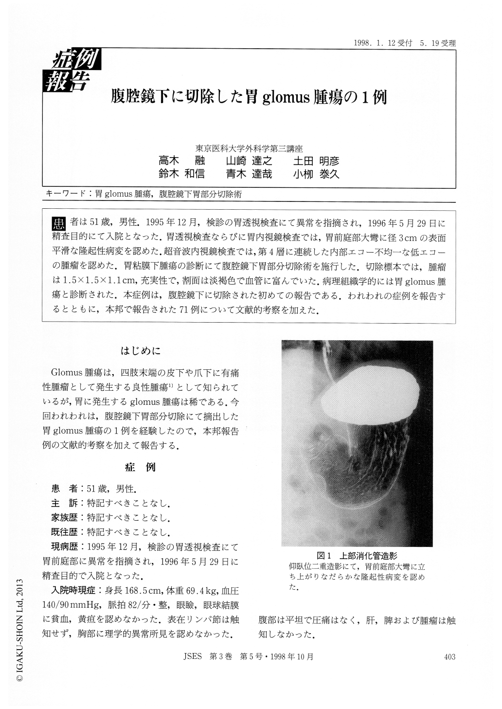
患者は51歳,男性.1995年12月,検診の胃透視検査にて異常を指摘され,1996年5月29日に精査目的にて入院となった.胃透視検査ならびに胃内視鏡検査では,胃前庭部大彎に径3cmの表面平滑な隆起性病変を認めた.超音波内視鏡検査では,第4層に連続した内部エコー不均一な低エコーの腫瘤を認めた.胃粘膜下腫瘍の診断にて腹腔鏡下胃部分切除術を施行した.切除標本では,腫瘤は1.5×1.5×1.1cm,充実性で,割面は淡褐色で血管に富んでいた.病理組織学的には胃glomus腫瘍と診断された.本症例は,腹腔鏡下に切除された初めての報告である.われわれの症例を報告するとともに,本邦で報告された71例について文献的考察を加えた.
A 51-year-old male was admitted to our department on May 29, 1996. Because of the abnormal findings detected by gastric fluoroscopy reformed in December 1995. Further fluoroscopic and gastroscopic examina-tions revealed a smooth raised lesion measuring 3cm in diameter, located on the greater curvature of the antrum. Endoscopic ultrasonography showed a heterogenous and hypoechoic mass which extended into the fourth layer. The lesion was diagnosed as submucosal tumor, and laparoscopic partial gastrectomy was performed.

Copyright © 1998, JAPAN SOCIETY FOR ENDOSCOPIC SURGERY All rights reserved.


