Japanese
English
- 有料閲覧
- Abstract 文献概要
- 1ページ目 Look Inside
- 参考文献 Reference
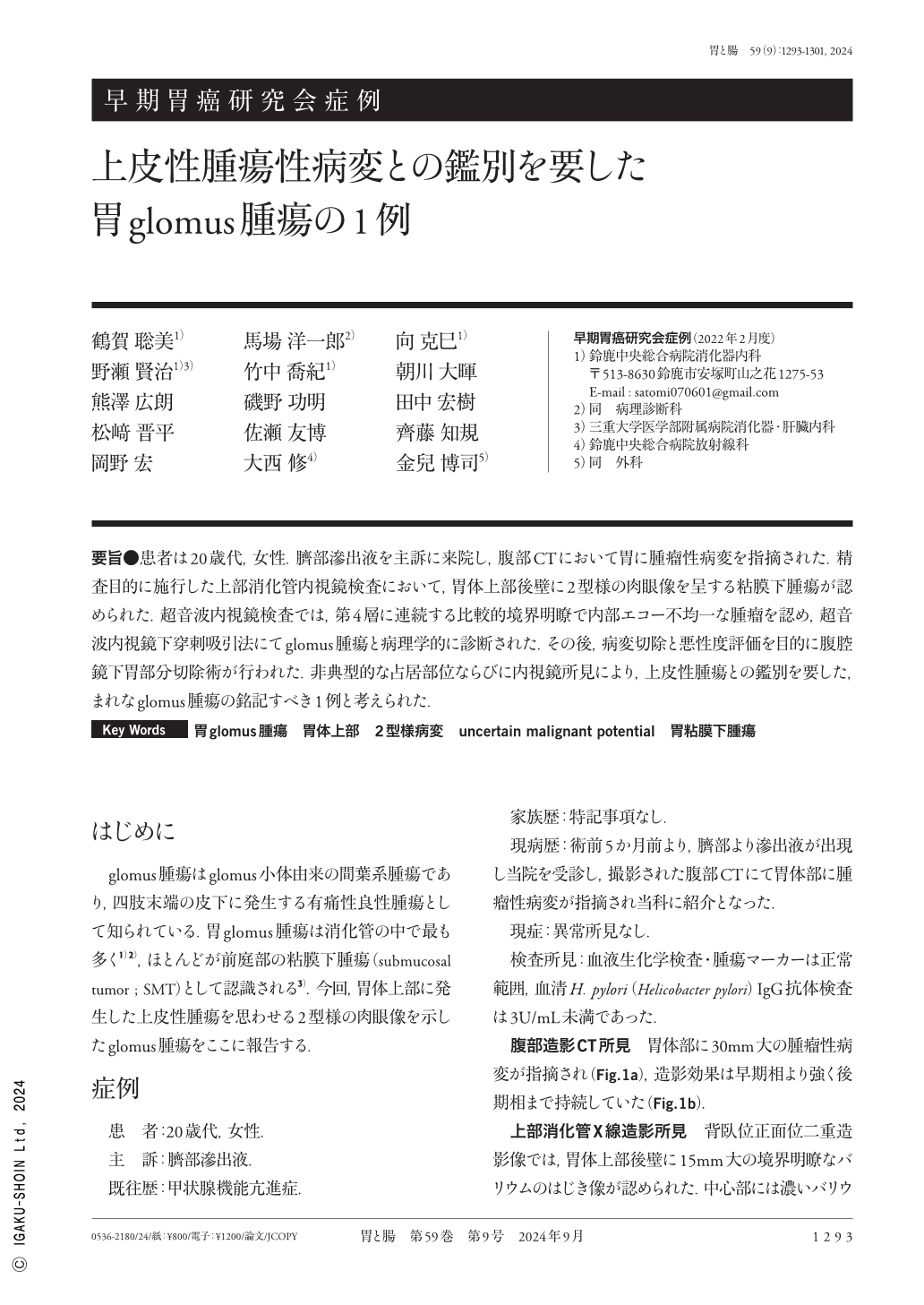
要旨●患者は20歳代,女性.臍部滲出液を主訴に来院し,腹部CTにおいて胃に腫瘤性病変を指摘された.精査目的に施行した上部消化管内視鏡検査において,胃体上部後壁に2型様の肉眼像を呈する粘膜下腫瘍が認められた.超音波内視鏡検査では,第4層に連続する比較的境界明瞭で内部エコー不均一な腫瘤を認め,超音波内視鏡下穿刺吸引法にてglomus腫瘍と病理学的に診断された.その後,病変切除と悪性度評価を目的に腹腔鏡下胃部分切除術が行われた.非典型的な占居部位ならびに内視鏡所見により,上皮性腫瘍との鑑別を要した,まれなglomus腫瘍の銘記すべき1例と考えられた.
A 20-year-old female presented with umbilical region effusion. Abdominal computed tomography detected a mass lesion in the stomach. Esophagogastroduodenoscopy demonstrated a submucosal tumor with a type 2-like structure on the posterior wall of the upper gastric body. Endoscopic ultrasonography(EUS)revealed a relatively well-defined mass with heterogeneous internal echogenicity in the fourth layer. On the basis of EUS-guided fine needle aspiration, the patient was diagnosed with a glomus tumor. Subsequently, a laparoscopic partial gastrectomy was performed to resect the lesion and assess its malignant potential. Herein, we describe a case of a glomus tumor originating from the upper gastric body that required differentiation from epithelial neoplastic lesions.

Copyright © 2024, Igaku-Shoin Ltd. All rights reserved.


