- 有料閲覧
- 文献概要
- 1ページ目
I.まえがき
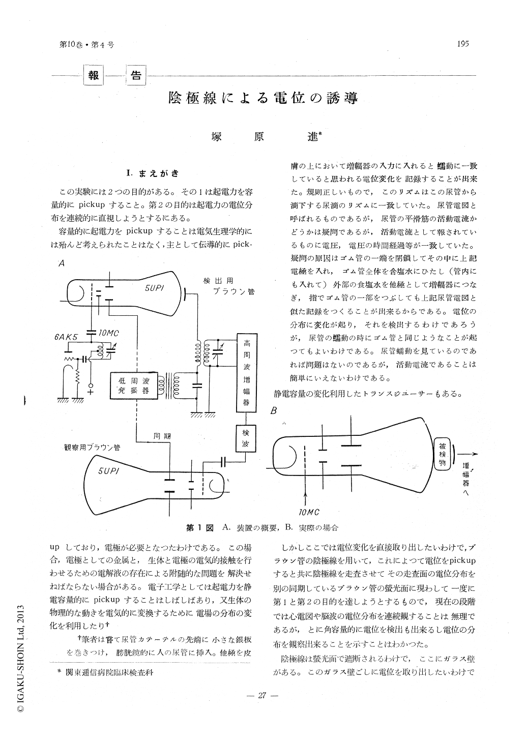
この実験には2つの目的がある。その1は起電力を容量的にpickupすること。第2の目的は起電力の電位分布を連続的に直視しようとするにある。
容量的に起電力をpickupすることは電気生理学的には殆んど考えられたことはなく,主として伝導的にpick-upしており,電極が必要となつたわけである。この場合,電極としての金属と,生体と電極の電気的接触を行わせるための電解液の存在による附随的な問題を解決せねばならない場合がある。電子工学としては起電力を静電容量的にpickupすることはしばしばあり,又生体の物理的な動きを電気的に変換するために電場の分布の変化を利用したり†
The author tried some experiments in order to obtain contur maps of electrical activity over the selected areas of the animal surface in rapid sequence. How to obtain such maps, C. Barus1), J. C. Lilly2) and other authors3) reported using many electrodes and amplifying systems which consisted of gate pulse generaters and switching circuits for each unit.
Instead of many electrodes and their amplifying systems, in this paper, a cathode ray tube was used as electrodes (electrodes tube), and the potential maps were illustrated on the screan of the other cathode ray tube (observation tube) as an array of illuminated spots, the brightness of each spot indicating the potential at a corresponding spot of the electrodes tube. The screan of the electrodes tube was set holded up to the surface having electrical uneveness and the beam was scanned the surface through the glass wall of the screan. The beam was chopped by 10 MC AC at the brightness control grid of the electrodes tube, so that the beam current, being modurated by the potential of the surface, was detected through the glass wall by means of a high frequency amplifyer.
The DC component of this amplifyed AC corresponds to the potentials at the spot where the electrrodes beam passed. When the brightness of the other cathode ray tube was controled by this potentials and the beams of both tube were deflected by a pair of oscillators output, then maps of electrical uneveness could be observed on the screan as bright and dark patterns.
Copyright © 1959, THE ICHIRO KANEHARA FOUNDATION. All rights reserved.