Japanese
English
- 有料閲覧
- Abstract 文献概要
- 1ページ目 Look Inside
1.緒言
網膜の視細胞外節中に存在する感光物質としては,桿体外節のRhodopsin及びPorphyropsinが詳細に研究され,その分光化学的性質も明らかにされている。一方錐体物質については,von Stüdnitz13),14),細谷・沖田・河久根8)以来種々の動物について研究が行われ,近くはWald15)17),Dartnall4)5)9)細谷・木村9)等の新らしい結果も発表されているが,錐体視物質として提唱されているもののλmaxは,近縁種の動物についても必らずしも一致していない。
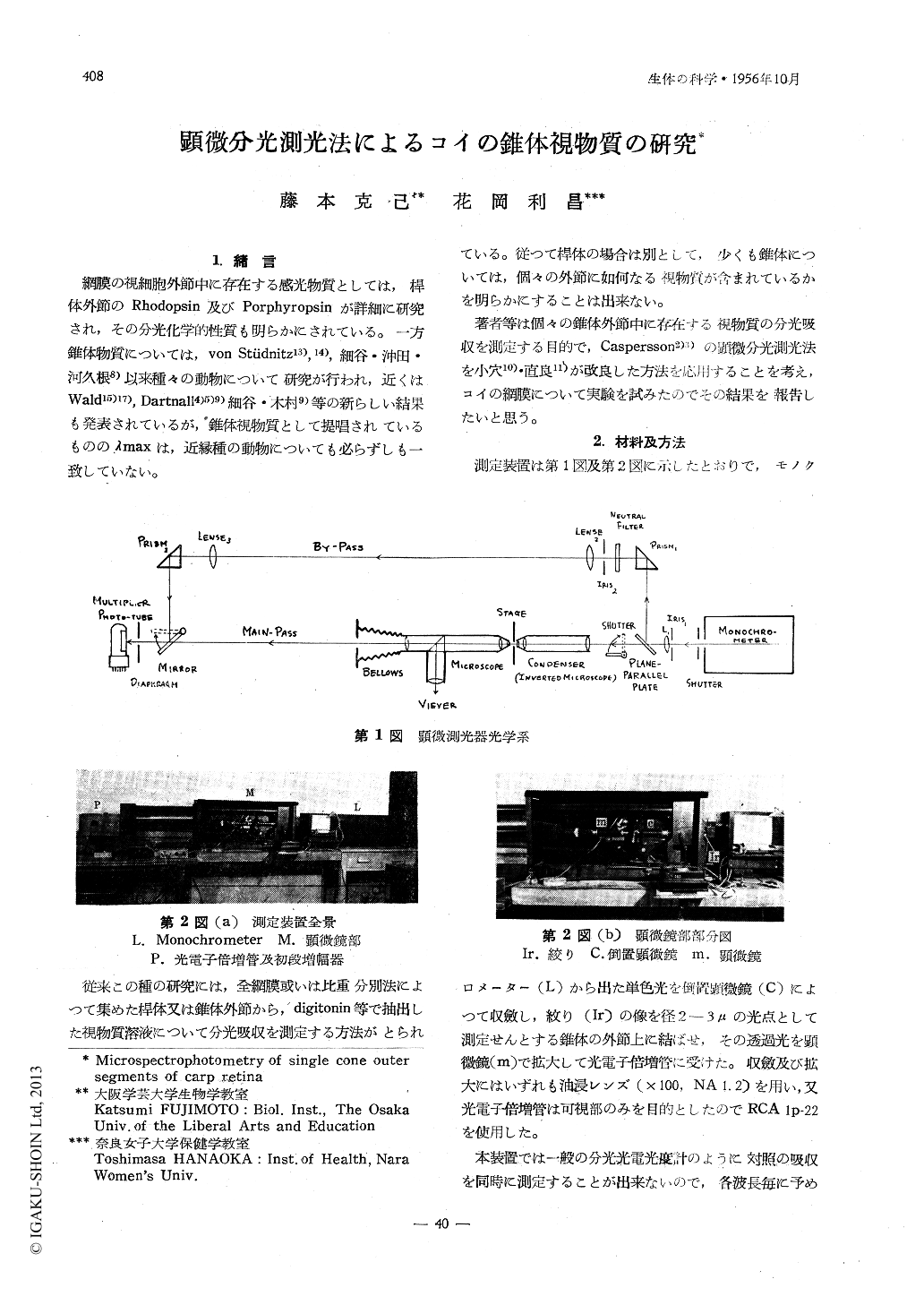
従来この種の研究には,全網膜或いは比重分別法によつて集めた桿体又は錐体外節から,digitonin等で抽出した視物質溶液について分光吸収を測定する方法がとられている。従つて桿体の場合は別として,少くも錐体については,個々の外節に如何なる視物質が含まれているかを明らかにすることは出来ない。
No one doubts the cone outer segments must contain photosensitive pigments like the rods do. Neyertheless, we can not detect these pigments in the extracted solution from the whole retina because of the dominancy of the rod pigment. The existence of the cone photosensitive pigments is only estimated in the so-called cone-retina in which the rod cells are very rare or almost absent. It is entirely impossible by the extraction method to verify whether the different photosensitive pigments are contained in every individual cone outer segments or not. In this case, the microspectrophotometry is the most ingeneous method.

Copyright © 1956, THE ICHIRO KANEHARA FOUNDATION. All rights reserved.


