Japanese
English
- 有料閲覧
- Abstract 文献概要
- 1ページ目 Look Inside
I.はじめに
特発性髄液鼻漏(essential cerebrospinal fluidrhinorrhea)はThomson1)により初めて記載されており,外傷や手術などに起因しない自然の髄液流出をいい,Hooper2)によれば1982年までに140例の報告をみている。本邦においても耳鼻咽喉科領域での報告が杉田ら3),野村ら4),青木ら5)などからなされており稀な疾患である。
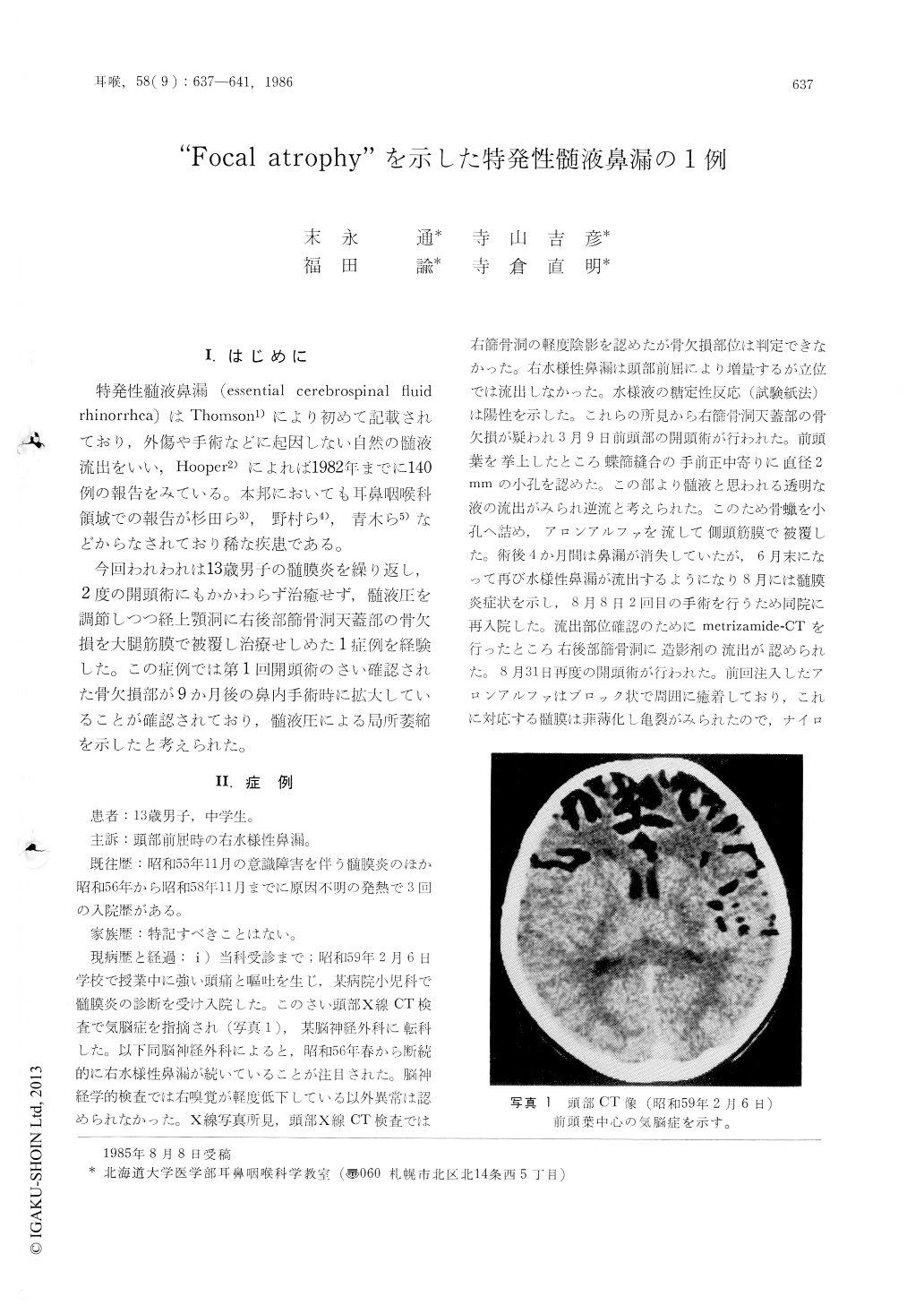
今回われわれは13歳男子の髄膜炎を繰り返し,2度の開頭術にもかかわらず治癒せず,髄液圧を調節しつつ経上顎洞に右後部筋骨洞天蓋部の骨欠損を大腿筋膜で被覆し治療せしめた1症例を経験した。この症例では第1回開頭術のさい確認された骨欠損部が9か月後の鼻内手術時に拡大していることが確認されており,髄液圧による局所萎縮を示したと考えられた。
A 13-year-old boy complaining of right watery rhinorrhea was suffered from repeated meningitis five times for four years.
Despite of two craniotomies, cerebrospinal rhinorrhea from the right ciribriform plate area was recurred. The bony defect had become larger than that at the first craniotomy, and it was thought to be "CSF blow out" proposed by Ommaya. Determination of the precise site of the CSF leak was made by metrizamide CT, plane x-ray and tomography. Antro-ethmoidectomy revealed a bony defect and meningocele at the tegmen of the right posterior ethmoid sinus. The defect was closed with fascia lata between dura and bone, and the other piece of fascia covered the defect. Before the operation spinal tap was made in order to make it easy to detect the leakage site by injection of saline, and to close the defect by aspiration of CSF. The dranage continued for seven days postoperatively.

Copyright © 1986, Igaku-Shoin Ltd. All rights reserved.


