Japanese
English
- 有料閲覧
- Abstract 文献概要
- 1ページ目 Look Inside
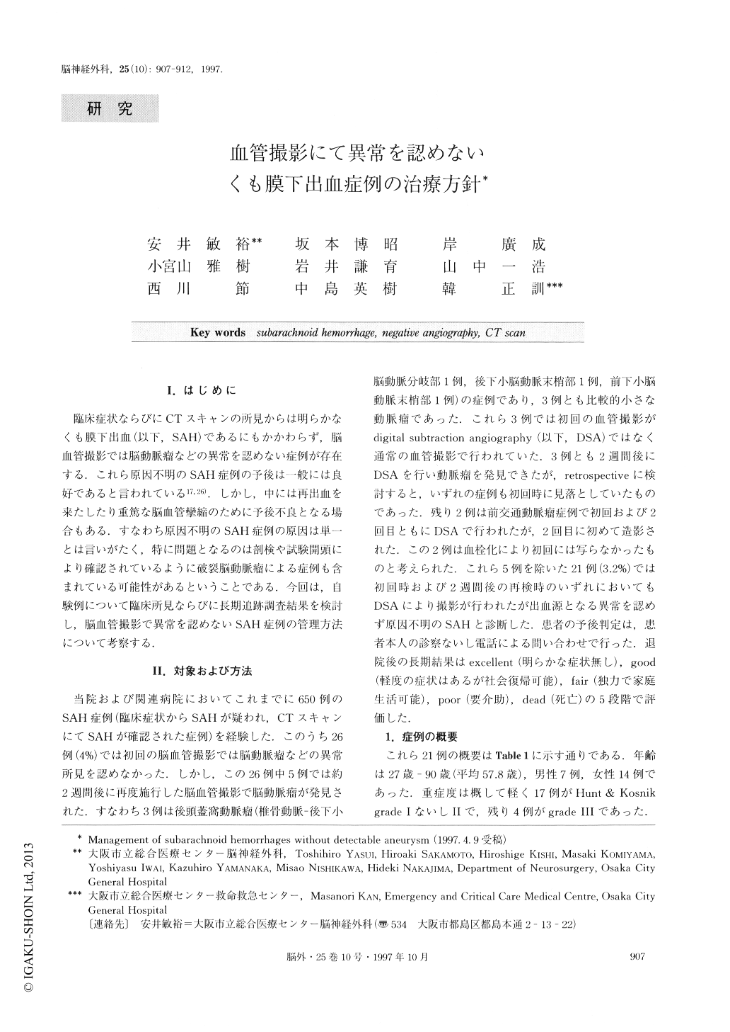
I.はじめに
臨床症状ならびにCTスキャンの所見からは明らかなくも膜下出血(以下,SAH)であるにもかかわらず,脳血管撮影では脳動脈瘤などの異常を認めない症例が存在する.これら原因不明のSAH症例の予後は一般には良好であると言われている17,26).しかし,中には再出血を来たしたり重篤な脳血管攣縮のために予後不良となる場合もある.すなわち原因不明のSAH症例の原因は単一とは言いがたく,特に問題となるのは剖検や試験開頭により確認されているように破裂脳動脈瘤による症例も含まれている可能性があるということである.今回は,自験例について臨床所見ならびに長期追跡調査結果を検討し,脳血管撮影で異常を認めないSAH症例の管理方法について考察する.
In this study, 21 patients with subarachnoid hemor-rhage (SAH) but negative angiography were evaluated. Angiography was performed twice on each patient, that is, on admission and at 2 weeks following admission. All patients had severe headache of sudden onset, a characteristic manifestation of SAH. Clinical grades on admission (Hunt and Kosnik classification) were gener-ally good: 17 patients were in grade Ⅰ or Ⅱ and 4 pa-tients were in grade Ⅲ. SAH was confirmed by the presence of subarachnoid clot on CT in all cases. Based on the distribution of SAH, CT findings were classified into two patterns, i. e., perimesencephalic and non-perimesencephalic patterns. Four patients showed the perimesencephalic pattern and the remaining 17 the non-perimesencephalic. The period of follow-up ranged from 20 days to 11 years 6 months, with a mean of 6 years 10 months. Except for three recent cases, themean follow-up period is 8 years 9 months. Exploratory craniotomies probing for aneurysms have been per-formed in four patients, but no aneurysms have been found in any of these cases. Clinical deterioration associated with vasospasm was observed in one patient. A communicating hydrocephalus requiring a shunting procedure was observed in three patients showing the non-perimesencephalic type CT pattern. Rebleeding occurred in one patient who subsequently died of what may be a dissecting aneurysm of the vertebral artery. One patient who was able to return to full activity ex-perienced symptoms attributable to SAH such as fre-quent headaches and increased fatigability. Complete recovery was observed in the remaining 19 patients. Two of them, however, later died due to myocardial in-farction and aging, respectively.
Given these generally positive outcomes, it should be possible to inform such patients of the benignity of their condition. Angiography may not demonstrate a ruptured aneurysm on initial examination in all cases of aneurysmal SAH. Serial angiography, however, can provide a definite diagnosis of the dissecting aneurysm. Therefore, repeat angiography, particularly, when possible, digital subtraction angiography, is necessary to rule out aneurysmal SAH. While small aneurysms or microaneurysms are often found through exploratory craniotomy, we do not agree with the opinion that surgery may be appropriate for certain patients with SAH but with negative angiography. The natural his-tory concerning rebleeding in such cases, as well as morbidity and mortality associated with hemorrhage, remains to be defined. Furthermore, there are reserva-tions regarding whether coagulation of these abnorma-lities with bipolar cautery constitutes definitive treat-ment.

Copyright © 1997, Igaku-Shoin Ltd. All rights reserved.


