Japanese
English
- 有料閲覧
- Abstract 文献概要
- 1ページ目 Look Inside
I.はじめに
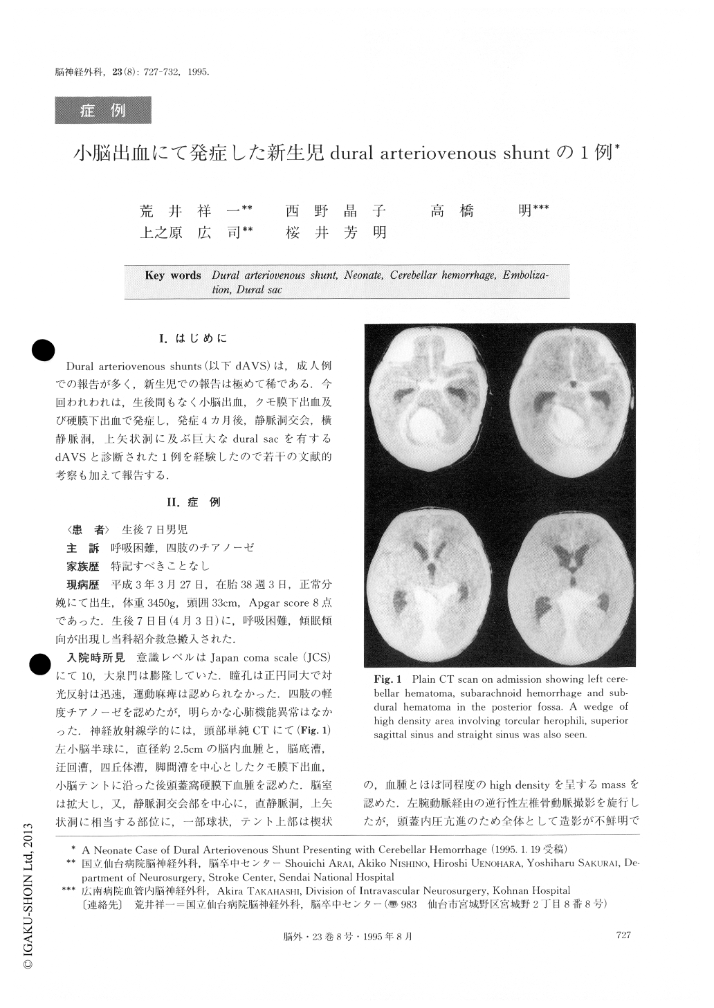
Dural arteriovenous shunts(以下dAVS)は,成人例での報告が多く,新生児での報告は極めて稀である.今回われわれは,生後間もなく小脳出血,クモ膜下出血及び硬膜下出血で発症し,発症4カ月後,静脈洞交会,横静脈洞,上矢状洞に及ぶ巨大なdural sacを有するdAVSと診断された1例を経験したので若千の文献的考察も加えて報告する.
A rare case of dural arteriovenous shunt (dAVS) manifested as cerebellar hemorrhage in a neonate is re-ported. Seven days after birth, a neonate was referred to our hospital because of consciousness disturbance. CT scan revealed cerebellar hemorrhage, subarachnoid hemorrhage, subdural hematoma, and a high density mass lesion at the torcular herophili. Retrograde brac-hial angiogram failed to show any vascular lesions. He underwent an evacuation of the cerebellar hematoma. Postoperative course was uneventful. However, at the age of four months, he was admitted again because of consciousness disturbance and cardiac failure. CT scan revealed hydrocephalus and an enlarged mass lesion at the torcular herophili.
Angiogram disclosed dural AVS. Its feeding arteries were as follows; the bilateral middle meningeal arte-ries (MMA), the occipital arteries (OA), dural branches of the bilateral posterior inferior cerebellar arteries, arteries consisting of transdural anastomosis from the left posterior cerebral artery. The arterial flow from these feeding arteries was shunting directly to the torcular herophili, the posterior part of the superior sagittal sinus, and the straight sinus. He underwent a venticulo-peritoneal shunting. Then, after treatment for cardiac failure, superselective embolization of the bi-lateral MMAs and the OAs resulted in diminution of shunting flow.
The initial onset of this case was at seven days after his birth. Dural AVS is very rare in the pediatric popu-lation, particularly in the neonate. The clinical features, pathogenesis, and the treatment for this rare entity are discussed.

Copyright © 1995, Igaku-Shoin Ltd. All rights reserved.


