Japanese
English
- 有料閲覧
- Abstract 文献概要
- 1ページ目 Look Inside
I.はじめに
外傷性気脳症は臨床上しばしばみうけられる病態であるが,空気が脳実質内に限局する脳内気脳症はかなりまれでありその報告も少ない1,6,10,13,14).従来より本症の発生には特異な機序が関わっていることが指摘されている8,9).今回,当施設において2例の脳内気脳症を経験したので報告し,本症に対する治療方針を中心に検討する.
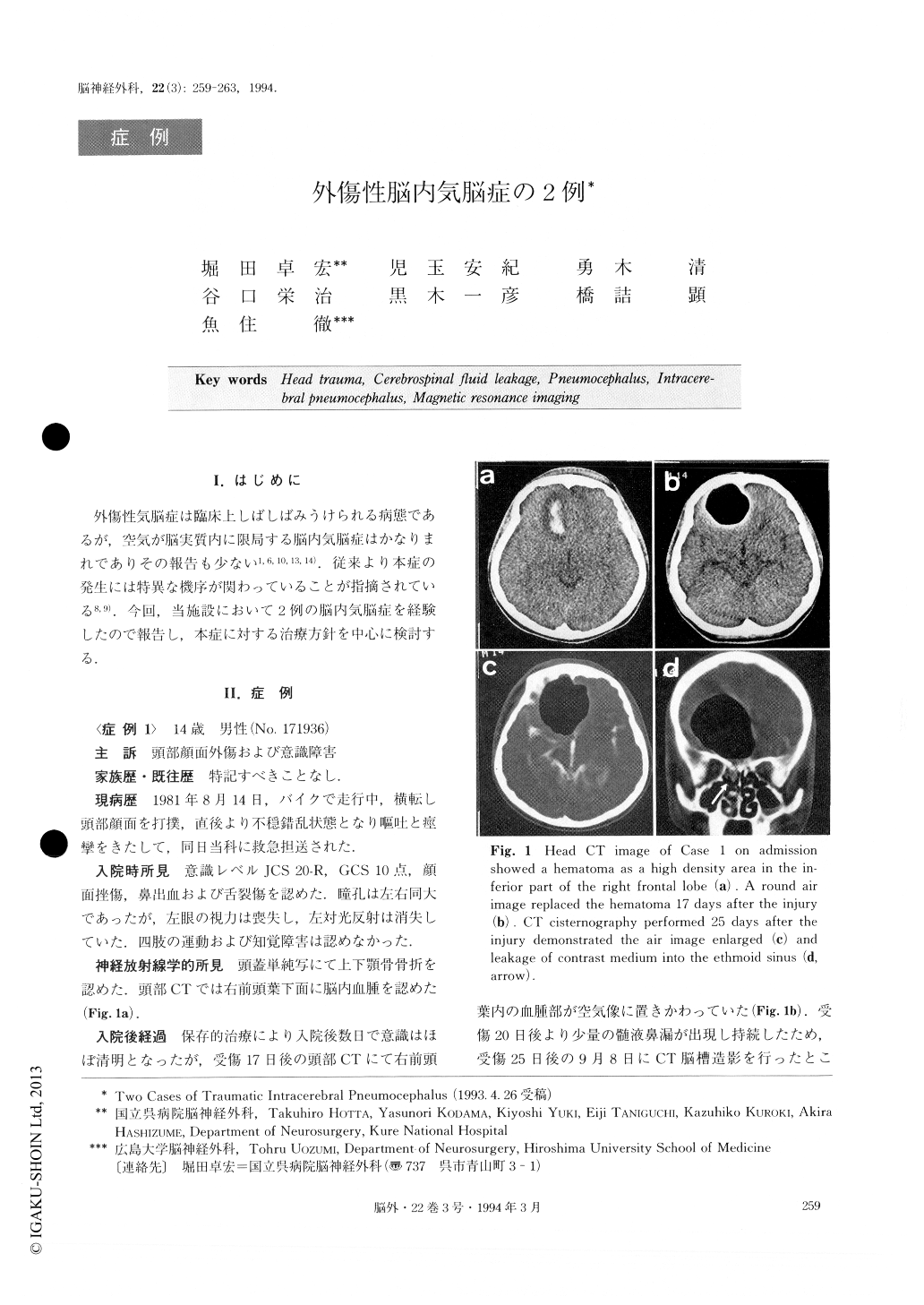
Two cases of traumatic intracerebral pneumocepha-lus, a rare complication of head trauma, are presented.Case 1:A 14-year-old boy had a strong concussion in his forehead due to a motorbike accident. Slightly obtunded on admission showing GCS 10, he became conscious in several days. Head CT performed after 17 days showed a round air image in the right frontal lobe which kept increasing in size thereafter. Bilateral fron-tal craniotomy was performed 31 days after the injury.A craniodural defect with a herniated brain was found in the superior wall of the posterior ethmoid sinus and repaired. Case 2: A 55-year-old man received a left forehead concussion when his motorbike ran into a car from behind. Although he had been conscious ever since admission, head CT after 15 days showed a round air image in the left frontal lobe. MRI demonstrated the air to be located in the cerebral parenchyma distinctly and the brain to have herniated into the frontal sinus. As the air showed a tendency to increase in volume and mild psychic and memory disturbances appeared, bi-lateral frontal craniotomy was performed 34 days after the injury. A craniodural defect with a herniated brain was detected in the posterior wall of the frontal sinus and repaired. These two patients showed a small amount of cerebrospinal fluid (CSF) rhinorrhea before the operation. Following the surgical repair, no recur-rence of pneumocephalus and CSF rhinorrhea has been seen in either case. Intracerebral pneumocephalus secondary to closed head trauma was thought to have been due to herniation of contused brain into a cra-niodural defect. For this reason, once intracerebral air volume increases, early surgical repair should be car-ried out for intracerebral pneumocephalus. Meticulous MRI investigation of the lesion causing intracerebral pneumocephalus is necessary for selection of an appropriate operative procedure.
Copyright © 1994, Igaku-Shoin Ltd. All rights reserved.