Japanese
English
- 有料閲覧
- Abstract 文献概要
- 1ページ目 Look Inside
I.はじめに
Rhinocerebrl phycomycosisは,Phycomycetes(藻菌)が鼻腔から副鼻腔を経て脳に浸潤病変をおこす稀な日和見真菌感染症で13,18),しばしば内頸動脈閉塞をもおこし致命的であることが多いといわれる1,18).本感染症は通常,コントロール不良な糖尿病や種々の免疫抑制状態や代謝異常疾患など18)の素因がみられるが,ごく稀には素因の不明な本感染症例が報告されている12,15,16).われわれは,素因の可能性として興味ある知見がみられた本感染症の手術治験例を経験したので文献的考察を加え報告する.
Rhinocerebral phycomycosis is an uncommon oppor-tunistic infection with ubiquitous fungi of the class Phycomycetes, starting in the nose and extending to the paranasal sinuses and then intracranially. The con-dition is often characterized by poor prognosis because of occlusion of the internal carotid artery. This diseasc is commonly associated with predispositions such as uncontrolled diabetes mellitus, which is the most com-mon, immunosuppressive states and metabolic bank-ruptcy including leukemia, lymphoma, myeloma, mal-nutrition, uremic or diarrheal acidosis, severe burns anemia, carcinoma, radiotherapy, liver cirrhosis, hemo-chromatosis, tuberculosis, septicemia, long-term medica-tion of steroid, antibiotics and antimetabolite, drug addiction, cytotoxic drug administration and AIDS.Cases with unknown predisposition, however, have been infrequently reported in the literature. The au-thors report a case of rhinocerebral phycomycosis in which concurrence of Candida species instead of the above-mentioned common predispositions was consi-dered a potential predisposition. To our knowledge, only 1 report in which Candida species are referred to as a potential predisposition for this disease has been previously issued.
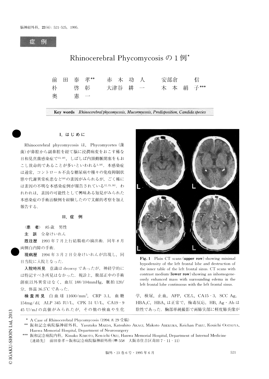
A 85-year-old man was admitted to our hospital on March 2, 1994 because of generalized convulsion. He had received a total extirpation of an ascending colon cancer in July 1993. On admission, physical inspectior showed no abnormalities and neurological examination revealed obtunded consciousness without other abnor-malities. He had no diabetes mellitus. Hematological and blood chemistry values were normal except for CA19-9 of 45U/ml. Cultures of sputa and pharyngeal swabs revealed no tubercle bacilli. Ultrasonics and CT of the abdomen showed no abnormalities. Bone and gallium scintigram of the whole body were also normal Skull X-ray films showed unclear superior margin and opaque upper portion of the left frontal sinus. CT of the brain revealed an inhomogeneously enhanced mass with surrounding edema in the left frontal lobe extend-ing into the left frontal sinus. MRI disclosed mucosal thickening of the left frontal sinus in addition to the mass shown on CT. Cerebral angiography was normal. A metastatic tumor was suspected judging from CA19-9 value and the patient's past history. Supraorbit-al craniectomy was performed. The tumor including the invaded dura and the thickened mucosa of the left fron-tal sinus was totally removed in a piecemeal manner us-ing microsurgical techniques. Histopathological diagno-sis was rhinocerebral phycomycosis. Culture for bacter-ia and fungi of cerebrospinal fluid taken a short period after definitive diagnosis was negative. But cultures of sputa and nasopharyngeal swabs taken in the same period yielded Candida species which were not de-tected preoperatively. Candida species did not dis-appear with administration of antifungal drug but did not produce clinical troubles thereafter. The paient left the hospital with no symptoms.
There have been reports of observations in vitro showing that Candidal peptide and polysaccharide mannan have deleterious effects on neutrophil chemotax-is and human phagocytic cell function. In the present case, therefore, concurrence of Candida species was thought to be a potential predisposition for this disease.

Copyright © 1995, Igaku-Shoin Ltd. All rights reserved.


