Japanese
English
- 有料閲覧
- Abstract 文献概要
- 1ページ目 Look Inside
I.はじめに
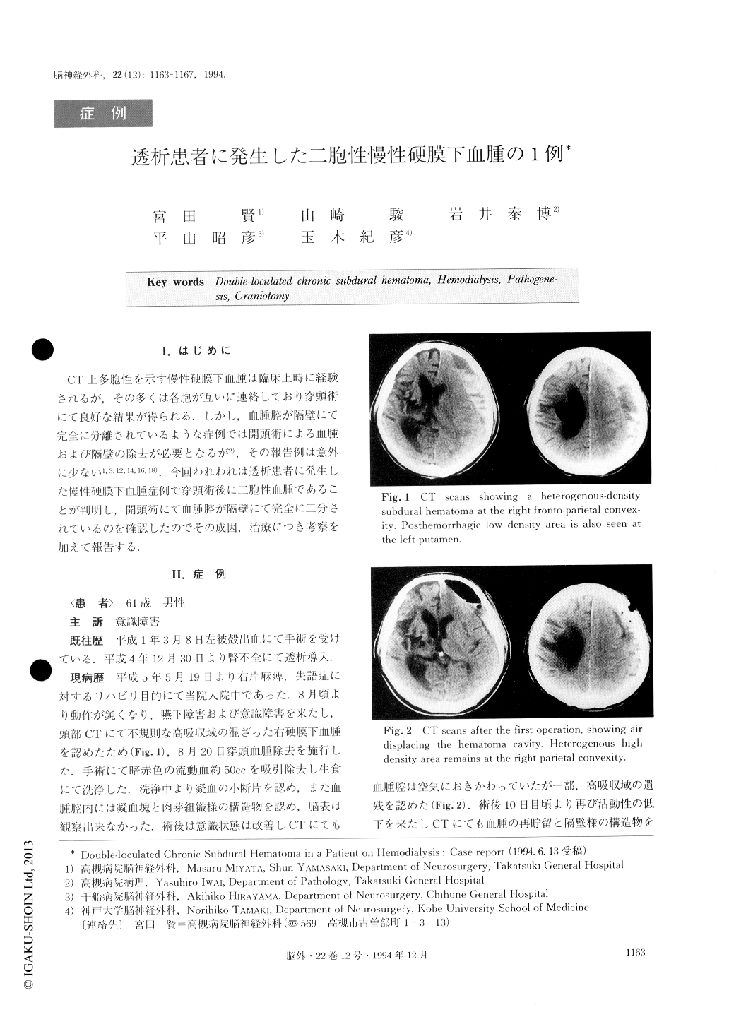
CT上多胞性を示す慢性硬膜下血腫は臨床上時に経験されるが,その多くは各胞が互いに連絡しており穿頭術にて良好な結果が得られる.しかし,血腫腔が隔壁にて完全に分離されているような症例では開頭術による血腫および隔壁の除去が必要となるが2),その報告例は意外に少ない1,3,12,14,16,18).今回われわれは透析患者に発生した慢性硬膜下血腫症例で穿頭術後に二胞性血腫であることが判明し,開頭術にて血腫腔が隔壁にて完全に二分されているのを確認したのでその成因,治療につき考察を加えて報告する.
A 61-year-old male who had been regularly hemo-dialyzed for a year and 8 months developed a double-loculated chronic subdural hematoma (CSDH). Com-puted tomography (CT) revealed a heterogenous-density hematoma at the right fronto-parietal convexity.After a burr hole aspiration and irrigation, CT dis-closed a residual hematoma and a septum separating the hematoma cavities. The hematoma and the septum were removed by right frontal craniotomy. At surgery, it was found that the thick septum completely sepa-rated the hematoma cavities and the outside hematoma was made up of coagula containing trabecula-like struc-tures, and the inside hematoma consisted of old bloody fluid.
Histological examination showed that the outer membrane and the septum were both thick because of the invasion by numerous lymphocytes which sug-gested that the intramembranous bleeding from sinu-soids split the outer membrane of an old CSDH. Double or multi-loculated CSDH should be consi-dered in a case which is slow in recovery or aggravated after a burr hole aspiration and irrigation.

Copyright © 1994, Igaku-Shoin Ltd. All rights reserved.


