Japanese
English
- 有料閲覧
- Abstract 文献概要
- 1ページ目 Look Inside
I.はじめに
硬膜動静脈奇形(DAVM)は横,S状静脈洞部に多発し過半数を占める20,21)とされ,大脳,小脳半球を覆う硬膜にDAVMが発生することは極めて稀で,そのほとんどは後頭蓋窩ないしは頭蓋底部に発生するといわれている.上矢状洞近傍の大脳半球円蓋部に発生した特発生DAVMの報告は文献上散見されるのみで,その部位的特異性は未だ明らかでなく,今回われわれの経験した1例を含め文献的考察を加えたのでここに結告する.
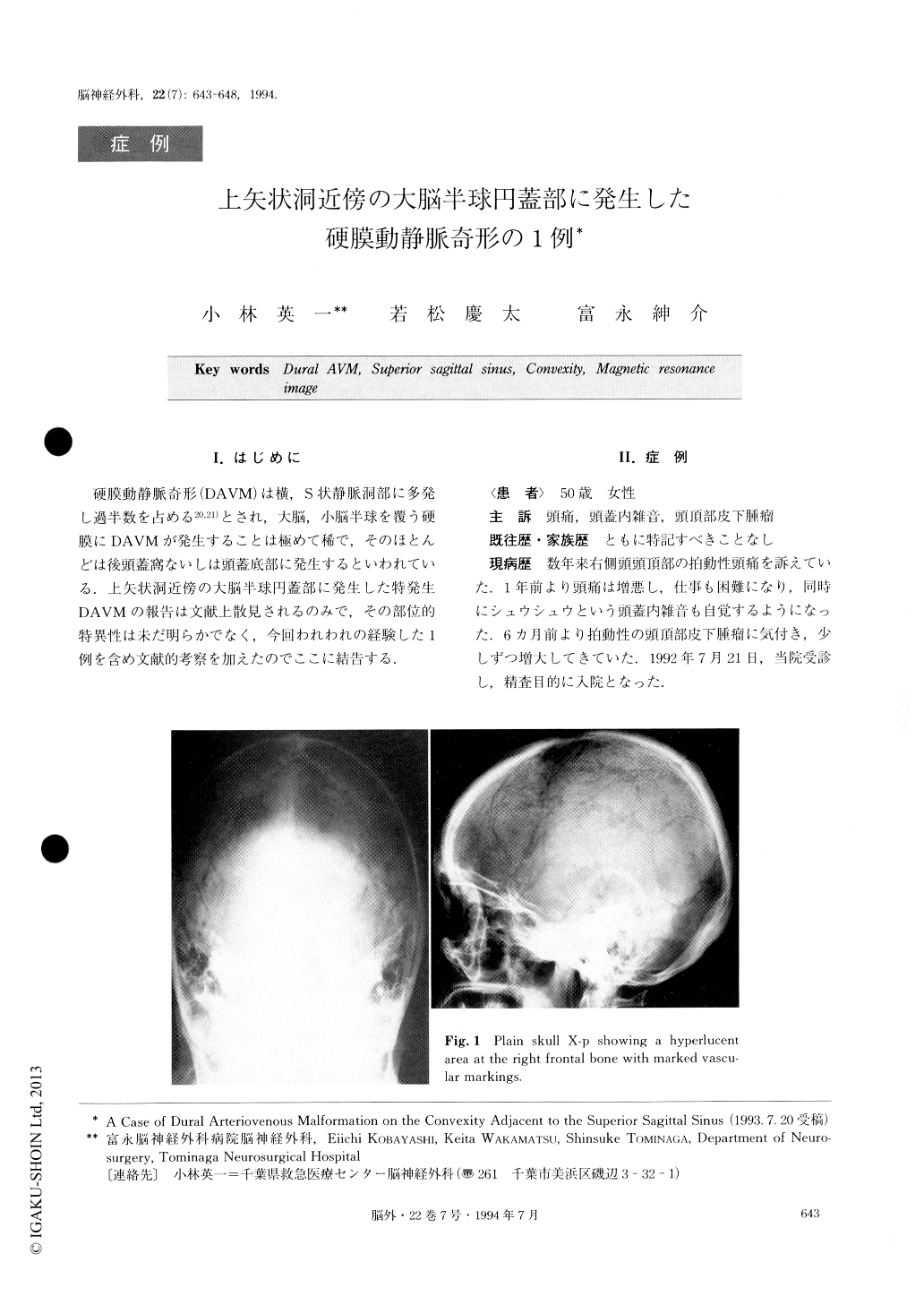
A case of chiral arteriovenous malformation (DAVM) on the convexity adjacent to the superior sagittal sinus (SSS) was reported. A 55-year-old female was admitted to our hospital complaining of severe headache and intracranial bruit. CT scan revealed an osteolytic lesion at the right frontal bone adjacent to the SSS, and MRI showed a flow void area at the same site. Cerebral angiography detected DAVM which was fed by bilateral superficial temporal arteries and plenty of meningeal branches of the middle meningeal artery. It was drained by cortical veins to the SSS. Endovascu-lar sugery was tried for this lesion, but it failed. After the surgical excision of DAVM, the above symptoms disappeared with no complications.
The 14 cases reported in the literature were ex-amined to characterize DAVM at this site. Average age was 47 with no age distribution. 43% of the cases had intracrnial hemorrhage, subarachnoid hemorrhage (2), intracranial hemorrhage (2), intraventricular hemor-rhage (1), and subdural hematoma (1). The DAVM arises most frequently at the middle third of the SSS and tends to extend to the posterior third. The symp-tom most frequently seen is headache, but we ought to pay attention to the possibility of progressive dementia.As for therapy, direct surgical excision is sometimes necessary if intravascular embolization ends in failure or incomplete cure.

Copyright © 1994, Igaku-Shoin Ltd. All rights reserved.


