Japanese
English
- 有料閲覧
- Abstract 文献概要
- 1ページ目 Look Inside
I.はじめに
硬膜動静脈瘻(奇形)は全頭蓋内動静脈奇形の約10%に発生するといわれ3),一般的にはテント下に多くテント上では大部分が海綿静脈洞部に発生する15,16).上矢状洞部に発生することはきわめて少なく流入動脈は両側性であることがほとんどである12,14).今回,われわれは脳内出血で発症した片側性上矢状静脈洞部の動静脈瘻を経験したので発生病態や臨床像を文献的考察を加えて報告する.
A case was reported of a surgically removed idiopathic dural arteriovenous fistula of the superior sagittal sinus associated with intracerebral hemorrhage. Dural arteriovenous fistula of the superior sagittal sinus is said to be rare. Only 9 cases have been reported in detail so far.
A 46-year-old male was referred to our clinic com-plaining of left motor weakness of sudden onset. He had no history of head injury.
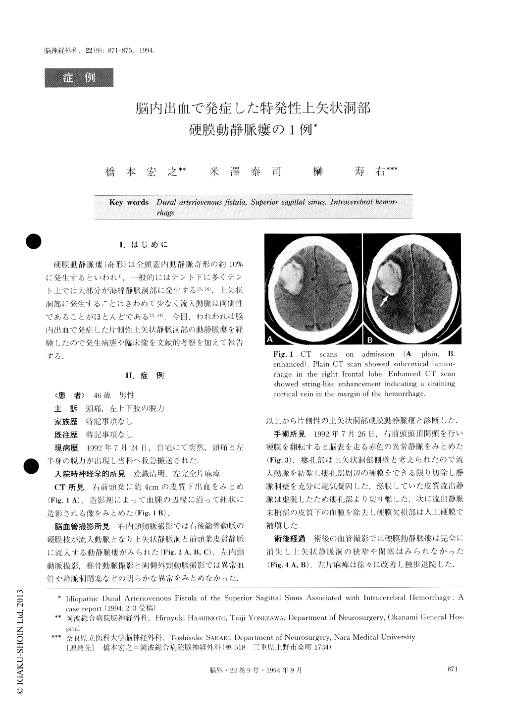
CT scan revealed subcortical hemorrhage in the right frontal lobe. Enhanced CT scan showed string-like en-hancement in the margin of the hemorrhage. Right in-ternal carotid angiogram disclosed a dural arteriove-nous fistula, of which the feeding artery was a mening-eal branch of the posterior ethmoidal artery, draining the superior sagittal sinus and right frontal superficial cortical vein. The superior sagittal sinus was not ob-structed. It was diagnosed as a rare idiopathic dural arteriovenous fistula in the superior sagittal sinus. Sur-gical obliteration of an arteriovenous fistula was per-formed preserving the superior sagittal sinus. Etiologic-al aspects and surgical management of the rare lesion was discussed referring to previous reports.

Copyright © 1994, Igaku-Shoin Ltd. All rights reserved.


