Japanese
English
- 有料閲覧
- Abstract 文献概要
- 1ページ目 Look Inside
I.はじめに
硬膜動静脈奇形(以下DAVM)は,横・S状静脈洞部および海綿静脈洞部に発生することが多く,大脳鎌DAVMの報告は非常に稀である.今回われわれは,くも膜下出血で発症した大脳鎌DAVMの1例を経験したので,若干の文献的考察を加えて報告する.
Abstract
A case of falx dural arteriovenous malformation was reported.
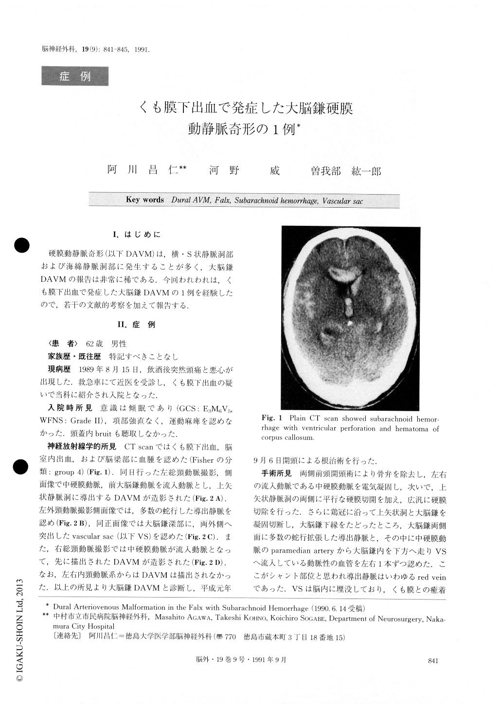
A 62 year old man was admitted to Nakamura City Hospital on August 15, 1989, with severe headache as his chief complaint. On admission, his consciousness was lethargic. CT scan showed subarachnoid hemor-rhage with ventricular perforation and hematoma of the corpus callosum. Angiograms demonstrated a dural arteriovenous malformation (DAVM) in the frontal falx, which was fed by bilateral middle meningeal arte-ries and the left anterior falx artery and drained into the superior sagittal sinus via the dural vein. Bifrontal craniotomy was performed. At first, bilateral middle meningeal arteries were coagulated, and the frontopa-rietal dura was excised widely. Then, the falx was cut at the crista galli. The DAVM was found in the falx, including a vascular sac embedded in the brain tissue. The DAVM was coagulated as much as possible. Caro-tid angiograms revealed complete disappearance of the DAVM, 4 months after the operation. Although angio-grams performed after only one month still showed a small residual DAVM.
On reviewing the literature we found only 5 patients with the DAVM in the falx. In 6 cases including our own, intracranial hemorrhage occurred in 4 cases (3 cases were subarachnoid hemorrhage). Vascular sacs were seen in 4 cases, and drainage to the pial vein was noted in 3 cases. It seemed to be rare that the DAVM drained into the dural vein. In our particular case, op-erative findings showed the DAVM drained into the dural vein without the pial vein, and intracranial hemorrhage was attributed to rupture of the vascular sac.

Copyright © 1991, Igaku-Shoin Ltd. All rights reserved.


