Japanese
English
- 有料閲覧
- Abstract 文献概要
- 1ページ目 Look Inside
I.はじめに
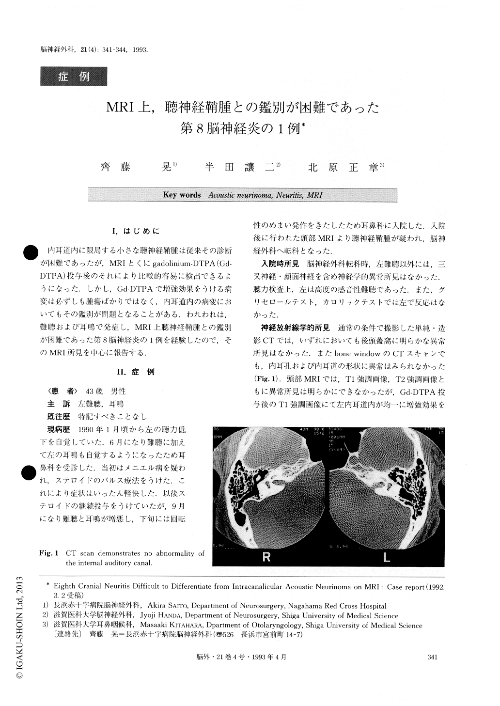
内耳道内に限局する小さな聴神経鞘腫は従来その診断が困難であったが,MRIとくにgadolinium-DTPA(Gd—DTPA)投与後のそれにより比較的容易に検出できるようになった.しかし,Gd-DTPAで増強効果をうける病変は必ずしも腫瘍ばかりではなく,内耳道内の病変においてもその鑑別が問題となることがある.われわれは,難聴および耳鳴で発症し,MRI上聴神経鞘腫との鑑別が困難であった第8脳神経炎の1例を経験したので,そのMRI所見を中心に報告する.
A patient with an enhancing, completely intracanalicu-lar mass on MRI was presented. He had noticed progres-sive hearing loss in the left ear with tinnitus. Neurologic-al examination revealed no abnormality except decreased hearing in the left ear. There were no other cranial nerve or cerebellar signs. An audiogram revealed pro-found hearing loss on the left ear with no ability of speech discrimination. Brainstem auditory evoked re-sponse was absent on the left. MRI enhanced with gadolinium-DTPA demonstrated an intracanalicular en-hancing lesion on the left which was presumed to be an intracanalicular acoustic neurinoma. The patient under-went a left suboccipital craniectomy. The eighth cranial nerve appeared normal in the cerebellopontine angle cist-ern, and was swollen and discolored in the internal audi-tory canal. It was removed piecemeal. The patient re-mained deaf in the left ear postoperatively. Histo-pathologically, the lesion consisted of edematous nerve fiber and inflammatory cells, but no tumor cell was pre-sent within the specimen. The patient was diagnosed as having neuritis. The clinical time course of symptoms in our patient was not unusual for an acoustic neurinoma. Itseems that the distinction between an intracanalicular acoustic neurinoma and other lesions cannot be made on basis of MR imaging alone. All available imaging modalities should be considered before a definitive sur-gical procedure is undertaken.

Copyright © 1993, Igaku-Shoin Ltd. All rights reserved.


