Japanese
English
- 有料閲覧
- Abstract 文献概要
- 1ページ目 Look Inside
I.はじめに
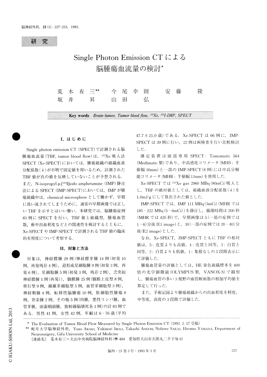
Single photon emission CT(SPECT)で計測される脳腫瘍血流量(TBF, tumor blood flow)は,133Xe吸入法SPECT(Xe-SPECT)においては,腫瘍組織の組織血液分配係数(λ)が不明で固定値を用いるため,計測されたTBF値が真の値を反映していないことが予想される.また,N-isopropyl-p—[123I]iodo amphetamine(IMP)静注法によるSPECT(IMP-SPECT)においては,IMPが腫瘍組織中は,chemical microsphereとして働かず,早期に洗い流されてしまうために,通常の早期画像では正しいTBFを示すとはいい難い.本研究では,脳腫瘍症例83例にSPECTを行い,TBF値と組織型,腫瘍血管数,術中出血程度などとの関連性を検討するとともに,Xe-SPECTやIMP-SPECTで計測されるTBF値の臨床的有用度について考察する.
The blood flow was measured in brain tumors (tumor blood flow: TBF). A total of 83 patients were studied. These included cases of 29 gliomas, 25 menin-giomas, 10 metastatic brain tumors and others. Measure-ments of TBF were performed using the 133Xe gas in-halation technique, the N-isopropyl-p-[123I] iodoampheta-mine intravenous injection method, and SPECT.
The results showed that TBF was variable, and was higher in meningioma. There was a significant correla-tion between TBF and the number of vessels in the op-erative specimen. Higher TBF tumors had a tendency to bleed profusely during operation. IMP El images were closely similar to Xe-SPECT images in TBF value. In cases with glioma, moderate correlation was noted between Xe-SPECT and IMP E2 image. A dis-crepancy in TBF values between IMP E2 image and Xe-SPECT image was observed. There was low acti-vity on IMP E2 image and high TBF value in Xe-SPECT image. This was true especially in cases with angioblastic meningioma.
In conclusion, TBF was considered to be helpful not only for preoperative diagnosis, but also for estimation of bleeding from tumors during operation and treat-ment

Copyright © 1993, Igaku-Shoin Ltd. All rights reserved.


