Japanese
English
- 有料閲覧
- Abstract 文献概要
- 1ページ目 Look Inside
I.はじめに
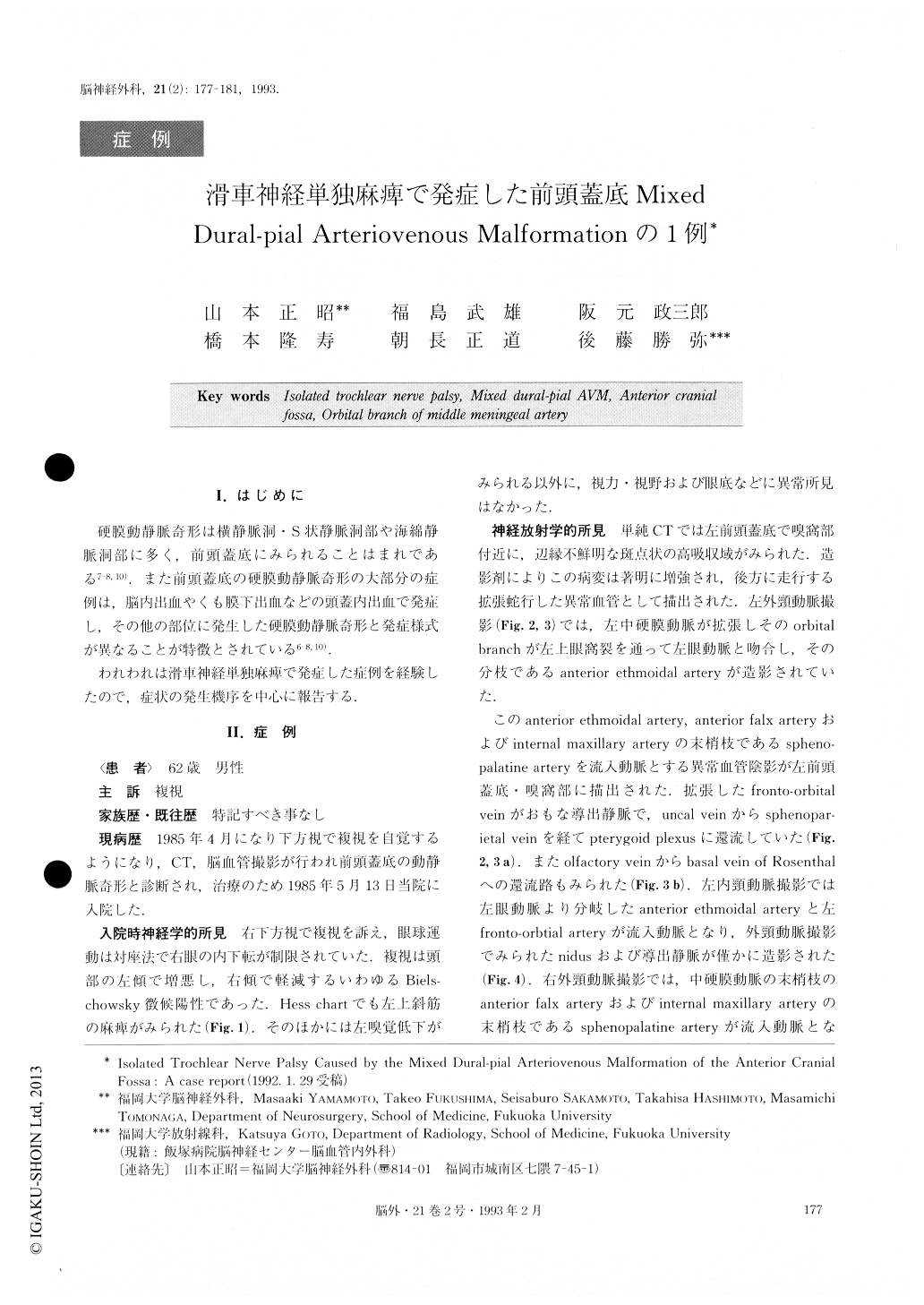
硬膜動静脈奇形は横静脈洞・S状静脈洞部や海綿静脈洞部に多く,前頭蓋底にみられることはまれである7-8,10).また前頭蓋底の硬膜動静脈奇形の大部分の症例は,脳内出血やくも膜下出血などの頭蓋内出血で発症し,その他の部位に発生した硬膜動静脈奇形と発症様式が異なることが特徴とされている6-8,10).
われわれは滑車神経単独麻痺で発症した症例を経験したので,症状の発生機序を中心に報告する.
A 62-year-old male was admitted to our hospital in May, 1985, with double vision, which had persisted for 1 month. The neurological examination revealed left troch-lear nerve palsy. A cerebral angiogram showed an arter-iovenous malformation at the anterior cranial fossa. The malformation was mainly fed by the left anterior ethmoidal artery which branched off from the orbital branch of the left middle meningeal artery. There was a marked enlargement of anastomosis between the orbital branch of the middle meningeal artery and the recurrent meningeal branch of the lacrimal artery. Bilateral distal branches of the internal maxillary arteries, bilateral anterior falx arteries and the left fronto-orbital artery were also involved in supplying the AVM. The dilated fronto-orbital vein was the main drainer which emptied into the pterygoid plexus via the uncal vein and the sphe-noparietal vein. In June 1985, the nidus involving the dura at the region of the cribriform plate and olfactory bulb was totally removed through a left frontal cra-niotomy. Postoperatively, the isolated left trochlear nerve palsy improved completely within a few days. This is the first reported case of isolated trochlear nerve palsy caused by a mixed dural-pial arteriovenous malformation of the anterior cranial fossa.
We concluded that the etiology of isolated trochlear nerve palsy consisted of nerve compression due to the di-lated orbital branch of the middle meningeal artery within the superior orbital fissure.

Copyright © 1993, Igaku-Shoin Ltd. All rights reserved.


