Japanese
English
- 有料閲覧
PPV購入案内
1,320円 (1,200円+税10%) こちらは、179ページから183ページの文献です。購入には医書.jp本会員登録が必要です。
会員登録完了後に改めて上記「購入サイトへ」を選択してください。
または「購入サイトへ」選択後に「購入ログイン」、「新規会員登録」の順に進んでください。
会員登録について詳しくはをご確認ください。
- Abstract 文献概要
- 1ページ目 Look Inside
I.はじめに
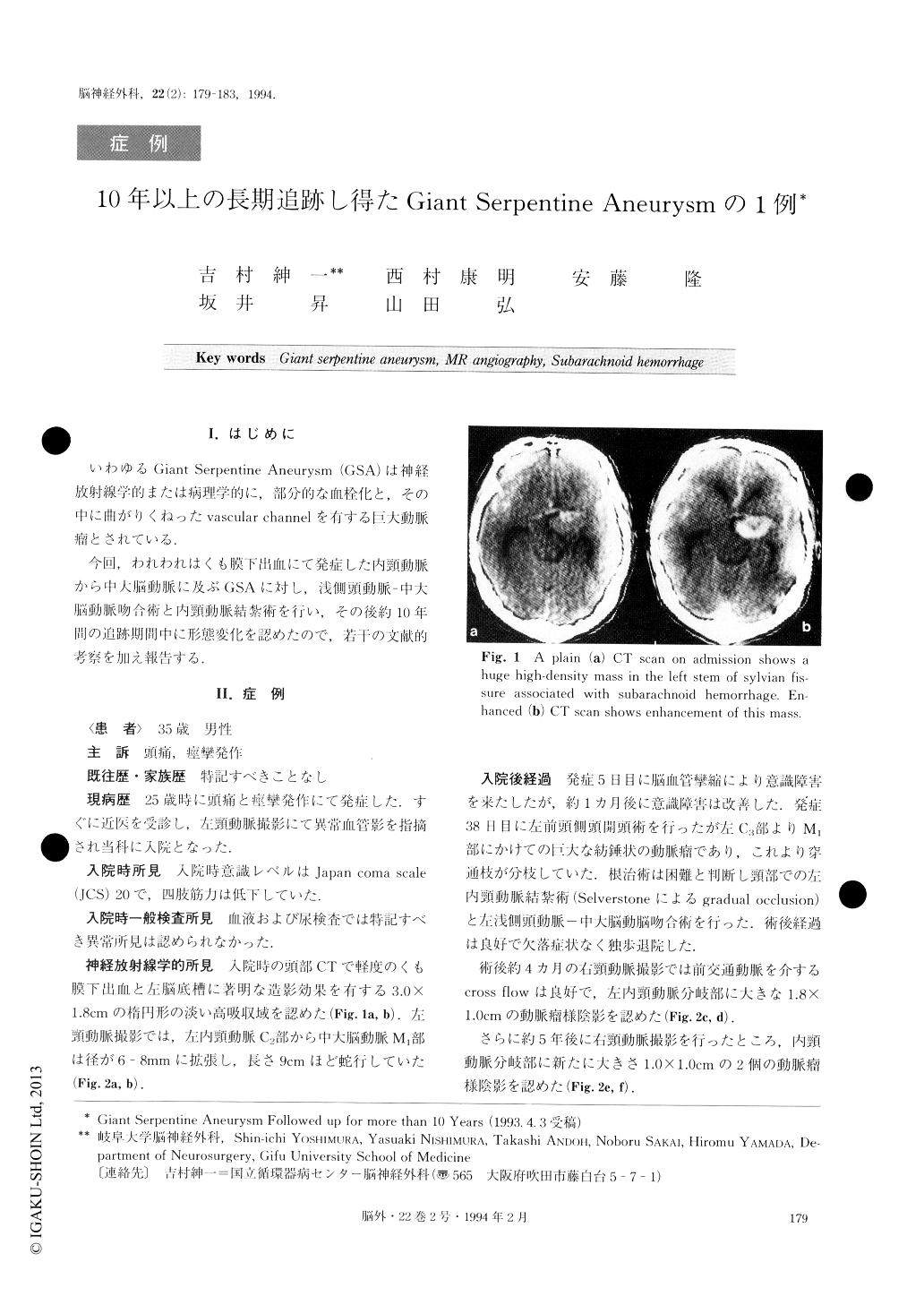
いわゆるGiant Serpentine Aneurysm(GSA)は神経放射線学的または病理学的に,部分的な血栓化と,その中に曲がりくねったvascular channelをる巨大動脈瘤とされている.
今回,われわれはくも膜下出血にて発症した内頸動脈から中大脳動脈に及ぶGSAに対し,浅側頸動脈—中大脳動脈吻合術と内頸動脈結紮術を行い,その後約10年間の追跡期間中に形態変化を認めたので,若干の文献的考察を加え報告する.
We report a case of a giant serpentine aneurysm (GSA) located at the left internal carotid artery (ICA) and middle cerebral artery (MCA) treated by ligation of the left ICA with superficial temporal artery-middle cerebral artery (STA-MCA) anastomosis. The aneu-rysm form changed variously during the follow-up period. A 35-year-old man was admitted with severe headache and convulsion. CT scan demonstrated sub-arachnoid hemorrhage. Left carotid angiogram demon-strated a giant serpentine aneurysm. Ligation of the ICA with STA-MCA anastomosis was performed be-cause of the difficulty involved in clipping. Right caro-tid angiogram obtained 5 years later revealed dis-appearance of the tortuous vascular channel and a new aneurysmal shadow at the left carotid bifurcation. The size of the aneurysmal shadow increased gradually over 10 years. MRI and MR angiography which clearly de-mostrated the presence of the aneurysm and the vascu-lar channel simultaneously were considered useful methods for diagnosis of GSA. The authors reviewed previous reports of 19 cases and investigated the mechanism of GSA.
Copyright © 1994, Igaku-Shoin Ltd. All rights reserved.