Japanese
English
- 有料閲覧
- Abstract 文献概要
- 1ページ目 Look Inside
I.はじめに
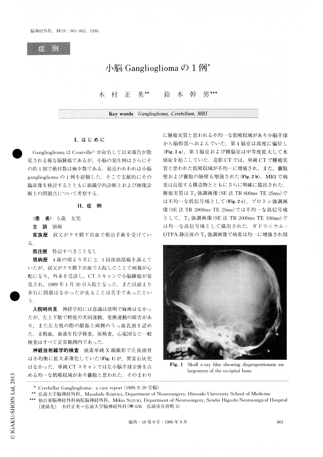
GangliogliomaはCourville1)が命名して以来報告が散見される稀な脳腫瘍であるが,小脳の発生例はさらにその約1割で絶対数は極少数である.最近われわれは小脳gangliogliomaの1例を経験した.そこで文献的にその臨床像を検討するとともに組織学的診断上および画像診断上の問題点について考察する.
A case of cerebellar ganglioglioma in a 5 year-old girl is presented. She came to our hospital on January 30, 1989 with complaints of headache of one year dura-tion. CT scans disclosed a low density lesion suggesting a cystic tumor in the left cerebellar hemisphere with moderate hydrocephalus. Preoperative MRI demon-strated more clearly the location and extent of the tumor.
She was operated on using suboccipital craniotomy, on March 3. Subtotal removal of the tumor was per-formed because the tumor had invaded the brain stem. She made an uneventful recovery without any neurolo-gical deficits. Histologically, the tumor was composed of ganglion cells and astrocytic cells, so it was di-agnosed as ganglioglioma.
Cerebellar ganglioglioma is a rare tumor, and only 17 cases have been reported including the present case. Clinical and radiological study of these cases revealed that there are no specific findings to indicate cerebellar ganglioglioma and preoperative diagnosis is impossible. But practically, MRI is the most sensitive method for identifying the extent of the lesion and, thus, is of be-nefit for deciding operative strategy.

Copyright © 1990, Igaku-Shoin Ltd. All rights reserved.


