Japanese
English
- 有料閲覧
- Abstract 文献概要
- 1ページ目 Look Inside
- 参考文献 Reference
Ⅰ.はじめに
大脳皮質下出血は,脳神経外科の日常臨床でよく遭遇する疾患である.本疾患では,血腫の脳への圧排による神経症状を改善する目的で,開頭血腫除去術が行われることがある.脳出血の原因は高血圧性が最も多いが,脳腫瘍からの出血の可能性も念頭に置かなければならない.
今回われわれは,左側頭葉皮質下出血に対し開頭血腫除去術を行い,術後1年で手術部位に膠芽腫が発生した症例を経験した.術後7カ月の時点で施行された頭部MRIでは腫瘍の所見を認めなかったため,脳出血の原因は腫瘍内出血よりも,手術後,脳出血手術部位に新たに発生した膠芽腫の可能性が考えられた.このような病態の存在は脳出血の術後経過観察方針に影響を与えると考えられるため,文献的考察を加えて報告する.
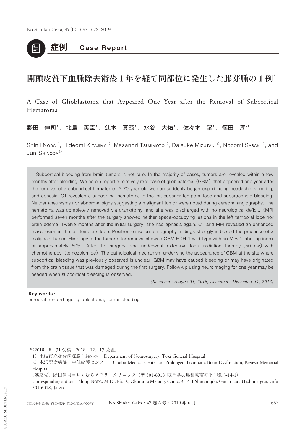
Subcortical bleeding from brain tumors is not rare. In the majority of cases, tumors are revealed within a few months after bleeding. We herein report a relatively rare case of glioblastoma(GBM)that appeared one year after the removal of a subcortical hematoma. A 70-year-old woman suddenly began experiencing headache, vomiting, and aphasia. CT revealed a subcortical hematoma in the left superior temporal lobe and subarachnoid bleeding. Neither aneurysms nor abnormal signs suggesting a malignant tumor were noted during cerebral angiography. The hematoma was completely removed via craniotomy, and she was discharged with no neurological deficit.(MRI performed seven months after the surgery showed neither space-occupying lesions in the left temporal lobe nor brain edema. Twelve months after the initial surgery, she had aphasia again. CT and MRI revealed an enhanced mass lesion in the left temporal lobe. Positron emission tomography findings strongly indicated the presence of a malignant tumor. Histology of the tumor after removal showed GBM HDH-1 wild-type with an MIB-1 labelling index of approximately 50%. After the surgery, she underwent extensive local radiation therapy(50 Gy)with chemotherapy(temozolomide). The pathological mechanism underlying the appearance of GBM at the site where subcortical bleeding was previously observed is unclear. GBM may have caused bleeding or may have originated from the brain tissue that was damaged during the first surgery. Follow-up using neuroimaging for one year may be needed when subcortical bleeding is observed.

Copyright © 2019, Igaku-Shoin Ltd. All rights reserved.


