Japanese
English
- 有料閲覧
PPV購入案内
1,320円 (1,200円+税10%) こちらは、921ページから926ページの文献です。購入には医書.jp本会員登録が必要です。
会員登録完了後に改めて上記「購入サイトへ」を選択してください。
または「購入サイトへ」選択後に「購入ログイン」、「新規会員登録」の順に進んでください。
会員登録について詳しくはをご確認ください。
- Abstract 文献概要
- 1ページ目 Look Inside
- 参考文献 Reference
Ⅰ.はじめに
脳内出血の原因としては高血圧症が最も頻度が高い6,13).高血圧性脳内出血は被殻,視床などの大脳基底核や歯状核(小脳)に発症することが多く,これらは穿通枝動脈の脆弱化に起因する一次性脳内出血に分類される.一方で,脳腫瘍の存在は,頻度は低いながらも脳出血の重要な原因の1つである4).特に,脳皮質下出血など高血圧性脳内出血の好発部位ではない場合には腫瘍の合併を念頭に置いておく必要がある.
今回われわれは,被殻出血で発症し保存的加療を行い,経過中の精査にて血腫とはやや離れた場所に脳腫瘍を認め,膠芽腫の診断に至った1例を経験したので文献的考察を含めて報告する.
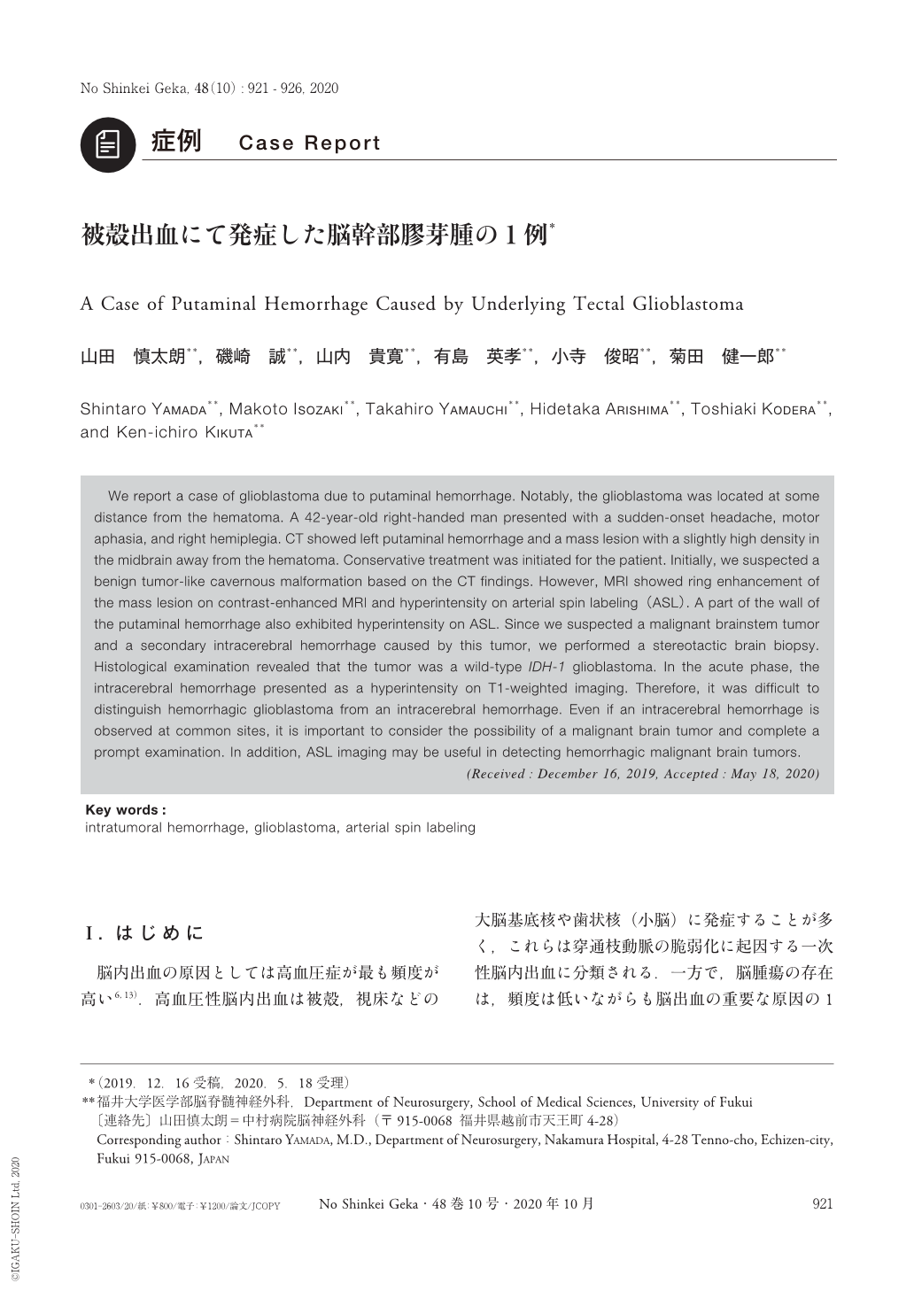
We report a case of glioblastoma due to putaminal hemorrhage. Notably, the glioblastoma was located at some distance from the hematoma. A 42-year-old right-handed man presented with a sudden-onset headache, motor aphasia, and right hemiplegia. CT showed left putaminal hemorrhage and a mass lesion with a slightly high density in the midbrain away from the hematoma. Conservative treatment was initiated for the patient. Initially, we suspected a benign tumor-like cavernous malformation based on the CT findings. However, MRI showed ring enhancement of the mass lesion on contrast-enhanced MRI and hyperintensity on arterial spin labeling(ASL). A part of the wall of the putaminal hemorrhage also exhibited hyperintensity on ASL. Since we suspected a malignant brainstem tumor and a secondary intracerebral hemorrhage caused by this tumor, we performed a stereotactic brain biopsy. Histological examination revealed that the tumor was a wild-type IDH-1 glioblastoma. In the acute phase, the intracerebral hemorrhage presented as a hyperintensity on T1-weighted imaging. Therefore, it was difficult to distinguish hemorrhagic glioblastoma from an intracerebral hemorrhage. Even if an intracerebral hemorrhage is observed at common sites, it is important to consider the possibility of a malignant brain tumor and complete a prompt examination. In addition, ASL imaging may be useful in detecting hemorrhagic malignant brain tumors.
Copyright © 2020, Igaku-Shoin Ltd. All rights reserved.