Japanese
English
- 有料閲覧
- Abstract 文献概要
- 1ページ目 Look Inside
I.はじめに
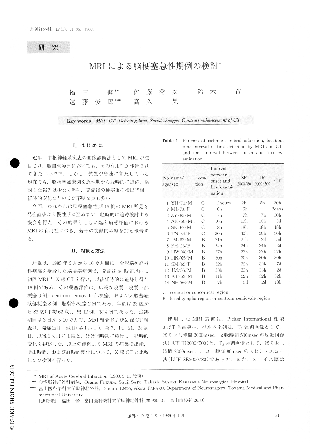
近年,中枢神経系疾患の画像診断法としてMRIが注目され,脳血管障害においても,その有用性が報告されてきた3-5,16,19,20).しかし,装置が急速に普及している現在でも,脳梗塞臨床例を急性期から経時的に追跡,検討した報告は少なく19,20),発症後の梗塞巣の検出時間,経時的変化などいまだ不明な点も多い.
今回,われわれは脳梗塞急性期16例のMRI所見を発症直後より慢性期に至るまで,経時的に追跡検討する機会を得た.その結果とともに臨床病態評価におけるMRIの有用性につき,若干の文献的考察を加え報告する.
Sequential changes of magnetic resonance imaging (MRI) in sixteen patients with acute cerebral infarction are studied in comparison with the findings of com-puted tomography (CT).
The sixteen patients were examined within 36 hoursfrom the onset of syptoms on resistive type MRI (0.15T) using T1 weighted image (1R2000/500) and T2 weighted image (SE2000/80), and on CT.
In general, large infarcted lesions of the cortex-subcortex seemed to be visualized earlier than small le-sions of the basal ganglia and brainstem. In 8 patients, the infarcted lesions were detected on MRI earlier than on CT.

Copyright © 1989, Igaku-Shoin Ltd. All rights reserved.


